Introduction
A goniometer is a device that measures an angle or permits the rotation of an object to a definite position. In orthopedics, the former description applies more. The art and science of measuring the joint ranges in each joint plane are called goniometry. Goniometry originates from 2 Greek words: gonia, which means angle, and metron, which means to measure. The first known use of a primitive version of the modern-day goniometer was by a Dutch physician and mathematician named Gemma Frisius, who used it to calculate and record the position of celestial bodies with respect to the earth.
Anatomy and Physiology
Register For Free And Read The Full Article
Search engine and full access to all medical articles
10 free questions in your specialty
Free CME/CE Activities
Free daily question in your email
Save favorite articles to your dashboard
Emails offering discounts
Learn more about a Subscription to StatPearls Point-of-Care
Anatomy and Physiology
The range of motion measures movement around a specific joint or body part. Doctors, osteopaths, physical therapists, or other health professionals commonly use a goniometer to measure the range of motion, an instrument that measures angle motion at a joint.[1][2] There are 3 types of range of motion, dependent on the purpose of the assessment: passive, active, and active assistive.
Types of Goniometers
Universal goniometers: These goniometers come in 2 forms: short-arm and long-arm. The short-arm goniometer is used for smaller joints like the wrist, elbow, or ankle. The long-arm goniometers are more accurate for joints with long levers like the knee and hip joints.[3] Of all the types, a universal goniometer is the most widely used.[4]
Twin axis electrogoniometer: The inter-rater and intra-rater reliability of the electrogoniometer is higher than that of the universal goniometer but challenging to apply in patients' clinical evaluation; hence, it is used more often for research purposes.[5]
Gravity goniometer/inclinometer: One arm has a weighted pointer that remains vertical under gravity.
Software/smartphone-based goniometer: A smartphone as a digital goniometer has several benefits, like availability, ease of measurement, application-based tracking of measurements, and one-hand use. These applications use the accelerometers in phones to calculate joint angles.[6][7][8][9]
Arthrodial goniometer: This goniometer is ideal for measuring cervical rotation, anteroposterior flexion, and lateral flexion of the cervical spine.
Indications
The goniometer is used in the following:
- Presence of dysfunction related to muscles, tendons, or joints
- Establishment of a diagnosis
- Development of treatment goals
- Evaluation progress or the lack of it
- Modification of treatment based on the progress
- Fabrication of orthoses
- Measurements for research purposes
Contraindications
Conditions for which a goniometer ought not to be used to measure active range of motion include the following:
- Joint dislocation
- Unhealed fracture
- Postsurgery, if movement disrupts the healing process
- Regions of osteoporosis or bone fragility, as forced measurements may cause iatrogenic injury
- Disruption of soft tissue likely following an injury
A goniometer is appropriate to use on the following conditions with added precautions:
- Infection or inflammation around a joint
- Severe pain aggravated by movement
- Hypermobility or instability
Equipment
A universal goniometer has 3 parts, which include the following:
- A body is designed like a protractor and may form a full or half-circle. It has a scale for the measurement of the angle. The scale can extend from 0 to 180 degrees for half-circle models or 0 to 360 degrees for full-circle models. The intervals on the scales can vary from 1 to 10 degrees.
- The fulcrum is a screw at the center of the body that allows the moving arm to move freely in the body of the device. The screw-like device can be tightened to fix the moving arm in a particular position or loosened to permit free movement. The fulcrum and the body are placed over the joint being measured.
- The stationary arm is the arm of the goniometer that aligns with the inactive part of the joint measured. It is structurally a part of the body and is not movable independently of the body. The moving arm is the arm of the goniometer, which aligns with the mobile part of the joint measured.
Another option is utilizing an accelerometer and rate gyroscope using a microelectromechanical system.
Personnel
Only trained physicians, doctors, physical therapists, occupational therapists, or other personnel with prior training should perform evaluations using goniometers.[10] This skilled person must know how to:
- Position and stabilize the joint correctly.
- Move a body part through its appropriate range of motion (ROM).
- Determine the joint's end range of motion, and end feel.
- Palpate the appropriate bony landmarks.
- Align the goniometer with the landmarks.
- Read the measuring instrument properly.
- Record measurements correctly.
Preparation
The use of a goniometer does not require elaborate preparation. The patient should be counseled well in advance, and consent for examination is necessary. The examination must be conducted in broad daylight, with the joint undergoing evaluation and the surrounding area well exposed. An assistant, if needed, should be called in advance.
Technique or Treatment
A goniometer can evaluate both active as well as passive range of motion. Positioning plays a vital part in goniometry because it helps to place the joints in a zero starting or neutral position and helps to stabilize the proximal joint segment. The examiner stabilizes the proximal joint component and then carefully moves the distal component of the joint through its entire available range of motion until reaching the end feel. After estimating the available range of motion, the examiner must conduct the following steps:
- Return the distal component to the starting position, palpate the relevant bony landmarks, and align the goniometer.
- Record the starting measurement, remove the goniometer, and allow the patient to move the joint through the available range of motion.
- Replace and realign the goniometer. Read and record the measurement.
- Repeat the measurement 3 times and calculate the average to obtain the active range of motion measurement.
- Compare the reading with the contralateral side.
- Move the joint passively through its passive range of motion (PROM) and repeat the abovementioned steps to measure PROM accurately.
- Care is necessary to ensure the patient does not move his body while moving the joint, ensuring accurate measurement.
Positioning significantly influences the tension in soft tissue structures like capsules, muscles, and ligaments, which envelope a joint. Any position that tenses the soft tissue structures leads to a limited range of motion compared to a position where the structures are lax.
It is vital to ensure that the same testing position is present during successive measurements to ensure that the amounts of tension remain constant in the soft tissue compared to past measurements. This approach assures the obtaining of similar results. Any change in position leads to erroneous readings.
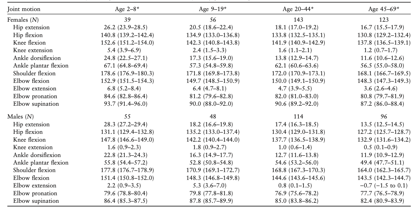
The range of motion differs from person to person by age and joint. Please see Table. Range of Motion According to Age and Joint.[11]
Complications
Complications related to goniometry are limited and are mainly due to faulty techniques; these include the following:
- Inaccurate measurement due to faulty technique can drastically affect the patient's treatment.
- Iatrogenic injuries in weak osteoporotic bones can result from forceful joint range of motion during goniometry.
Clinical Significance
Goniometric measurements can be useful in a variety of clinical scenarios. They range from mapping the spine mobility in cases of ankylosing spondylitis to checking the spine's range of motion after fusion surgeries for scoliosis. Improvements in the range of motion of the extremity joints can be easily noted with goniometric testing.
The overall consensus is still unsure whether or not the goniometer is a sufficiently valid and reliable device to know the effectiveness of an intervention.[12] The reliability of the results obtained from the goniometer might have a bearing on the type of goniometer used. There are also cases where a statistically significant difference is not observed.[13]
Enhancing Healthcare Team Outcomes
A goniometer can help in clinical decision-making regarding the management and outcome analysis after a particular intervention has been applied and compare the efficacies of different treatments. This methodology helps healthcare professionals identify the most efficacious treatment modality for a specific condition. Using goniometry results in maximized and enhanced patient outcomes.[14]
Media
References
Gates DH, Walters LS, Cowley J, Wilken JM, Resnik L. Range of Motion Requirements for Upper-Limb Activities of Daily Living. The American journal of occupational therapy : official publication of the American Occupational Therapy Association. 2016 Jan-Feb:70(1):7001350010p1-7001350010p10. doi: 10.5014/ajot.2016.015487. Epub [PubMed PMID: 26709433]
Keogh JWL, Cox A, Anderson S, Liew B, Olsen A, Schram B, Furness J. Reliability and validity of clinically accessible smartphone applications to measure joint range of motion: A systematic review. PloS one. 2019:14(5):e0215806. doi: 10.1371/journal.pone.0215806. Epub 2019 May 8 [PubMed PMID: 31067247]
Level 1 (high-level) evidenceHancock GE, Hepworth T, Wembridge K. Accuracy and reliability of knee goniometry methods. Journal of experimental orthopaedics. 2018 Oct 19:5(1):46. doi: 10.1186/s40634-018-0161-5. Epub 2018 Oct 19 [PubMed PMID: 30341552]
Nizamis K, Rijken NHM, Mendes A, Janssen MMHP, Bergsma A, Koopman BFJM. A Novel Setup and Protocol to Measure the Range of Motion of the Wrist and the Hand. Sensors (Basel, Switzerland). 2018 Sep 25:18(10):. doi: 10.3390/s18103230. Epub 2018 Sep 25 [PubMed PMID: 30257521]
Bronner S, Agraharasamakulam S, Ojofeitimi S. Reliability and validity of electrogoniometry measurement of lower extremity movement. Journal of medical engineering & technology. 2010 Apr:34(3):232-42. doi: 10.3109/03091900903580512. Epub [PubMed PMID: 20180734]
Ockendon M, Gilbert RE. Validation of a novel smartphone accelerometer-based knee goniometer. The journal of knee surgery. 2012 Sep:25(4):341-5 [PubMed PMID: 23150162]
Level 1 (high-level) evidenceJones A, Sealey R, Crowe M, Gordon S. Concurrent validity and reliability of the Simple Goniometer iPhone app compared with the Universal Goniometer. Physiotherapy theory and practice. 2014 Oct:30(7):512-6. doi: 10.3109/09593985.2014.900835. Epub 2014 Mar 25 [PubMed PMID: 24666408]
Ferriero G, Vercelli S, Sartorio F, Muñoz Lasa S, Ilieva E, Brigatti E, Ruella C, Foti C. Reliability of a smartphone-based goniometer for knee joint goniometry. International journal of rehabilitation research. Internationale Zeitschrift fur Rehabilitationsforschung. Revue internationale de recherches de readaptation. 2013 Jun:36(2):146-51. doi: 10.1097/MRR.0b013e32835b8269. Epub [PubMed PMID: 23196790]
Ferriero G, Sartorio F, Foti C, Primavera D, Brigatti E, Vercelli S. Reliability of a new application for smartphones (DrGoniometer) for elbow angle measurement. PM & R : the journal of injury, function, and rehabilitation. 2011 Dec:3(12):1153-4. doi: 10.1016/j.pmrj.2011.05.014. Epub [PubMed PMID: 22192326]
Carley P, Burkhart KL, Sheridan C. Virtual Reality vs Goniometry: Intraclass Correlation Coefficient to Determine Inter-Rater Reliability for Measuring Shoulder Range of Motion. Journal of allied health. 2021 Summer:50(2):161-165 [PubMed PMID: 34061937]
Soucie JM, Wang C, Forsyth A, Funk S, Denny M, Roach KE, Boone D, Hemophilia Treatment Center Network. Range of motion measurements: reference values and a database for comparison studies. Haemophilia : the official journal of the World Federation of Hemophilia. 2011 May:17(3):500-7. doi: 10.1111/j.1365-2516.2010.02399.x. Epub 2010 Nov 11 [PubMed PMID: 21070485]
Milanese S, Gordon S, Buettner P, Flavell C, Ruston S, Coe D, O'Sullivan W, McCormack S. Reliability and concurrent validity of knee angle measurement: smart phone app versus universal goniometer used by experienced and novice clinicians. Manual therapy. 2014 Dec:19(6):569-74. doi: 10.1016/j.math.2014.05.009. Epub 2014 Jun 4 [PubMed PMID: 24942491]
Mourcou Q, Fleury A, Diot B, Franco C, Vuillerme N. Mobile Phone-Based Joint Angle Measurement for Functional Assessment and Rehabilitation of Proprioception. BioMed research international. 2015:2015():328142. doi: 10.1155/2015/328142. Epub 2015 Oct 25 [PubMed PMID: 26583101]
Gajdosik RL, Bohannon RW. Clinical measurement of range of motion. Review of goniometry emphasizing reliability and validity. Physical therapy. 1987 Dec:67(12):1867-72 [PubMed PMID: 3685114]
