Introduction
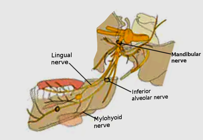
The trigeminal nerve (CN V) is responsible for sensory innervation of the face. The trigeminal nerve splits off into three main branches. The three branches that originate from the trigeminal nerve are the ophthalmic nerve (CN V1), the maxillary nerve (CN V2), and the mandibular nerve (CN V3). These nerves will provide sensory innervation to their respective territories. The trigeminal nerve also provides motor innervation to the muscles used in mastication. The maxillary nerve and the mandibular nerve will provide motor innervation to the muscles used for mastication. Each muscle that is involved in mastication receives a nerve that branches from the maxillary nerve or the mandibular nerve. For example, the mylohyoid muscle contributes to chewing, speaking, and swallowing. This muscle receives innervation from a nerve called the mylohyoid nerve. The mylohyoid nerve is one of the branches of the inferior alveolar nerve (branch of the mandibular nerve).
Structure and Function
Register For Free And Read The Full Article
Search engine and full access to all medical articles
10 free questions in your specialty
Free CME/CE Activities
Free daily question in your email
Save favorite articles to your dashboard
Emails offering discounts
Learn more about a Subscription to StatPearls Point-of-Care
Structure and Function
The mylohyoid nerve is a branch from the inferior alveolar nerve. The inferior alveolar nerve originates from the mandibular branch of the trigeminal nerve. The branching of the inferior alveolar nerve into the mylohyoid nerve occurs before the inferior alveolar nerve enters the mandibular foramen. Once the mylohyoid nerve branches, it will descend on the medial side of the mandibular ramus. The mylohyoid nerve will perforate through the sphenomandibular ligament to enter the mylohyoid groove and continues to descend towards the mylohyoid muscle. The mylohyoid nerve travels closely with the medial division of the submandibular glands, ultimately settling between the mylohyoid muscle and the anterior muscle belly of the digastric muscle. The anatomical position of the mylohyoid nerve is inferolateral to the mylohyoid muscle but superior to the anterior muscle belly of the digastric muscle.[1][2][3]
The mylohyoid nerve's function is to provide motor innervation to the mylohyoid muscle and the anterior muscle belly of the digastric muscle.[1] It also gives off a cutaneous nerve. This cutaneous nerve branch will provide sensory innervation to the inferior aspect of the chin. The mylohyoid nerve also provides sensory nerves to the first molar's mesial root.
Embryology
During fetal development, the structures in the face and neck form from the brachial apparatus. The brachial apparatus is made up of the brachial clefts, the brachial arches, and the brachial pouches. The brachial arches are responsible for developing the muscles, nerves, and bones. The brachial arches are made up of neural crest cells and mesoderm. The mesoderm will develop into the muscles and some of the bones while the neural crest cells will develop into the peripheral nerves and some of the facial bones. The maxillary nerve and the mandibular nerve branches of the trigeminal nerve are derivatives of the first brachial arch. As the mandibular nerve continues to develop and elongate, it will branch and form the inferior alveolar nerve as one of its branches. The inferior alveolar nerve will branch into the mylohyoid nerve, making the mylohyoid nerve a derivative of neural crest cells in the first brachial arch.
Blood Supply and Lymphatics
The blood supply to the mylohyoid nerve is predominately from the branches of the external carotid artery. The mylohyoid nerve receives perfusion from different arteries as it descends toward the mylohyoid muscle. The inferior alveolar arteries will perfuse the mylohyoid nerve around the mandible region. Once the mylohyoid nerve reaches the mylohyoid muscle, it will receive that same perfusion as the mylohyoid muscle. The arteries that perfuse the mylohyoid muscle are the mylohyoid branch of the inferior alveolar artery and the submental branches of the facial artery.[1]
The lymphatic drainage of the territory innervated by the mylohyoid nerve gets directed towards the submental lymph nodes and the submandibular lymph nodes. The lymph fluid will eventually drain toward the right lymphatic duct or the thoracic duct. The right lymphatic duct will drain the right side of the face and neck. The left side of the face and neck will drain toward the thoracic duct. The right lymphatic duct and the thoracic duct will drain back into the central circulation.[4]
Nerves
The mylohyoid nerve is a branch of the inferior alveolar nerve. The inferior alveolar nerve originates from the mandibular branch of the trigeminal nerve. The mylohyoid nerve provides both motor and sensory innervation.[3]
Muscles
The mylohyoid nerve will provide motor innervation to the mylohyoid muscle and the anterior muscle belly of the digastric muscle.
Physiologic Variants
The mylohyoid nerve is a nerve with many different variations. The length of the mylohyoid may vary depending on the origin of its branch point and the path of the nerve. The mylohyoid nerve is classically a branch of the inferior alveolar nerve, but in some instances, the mylohyoid nerve is a direct branch from the mandibular nerve. The path of the mylohyoid nerve usually occurs in the mylohyoid groove, but the nerve can descend toward the mylohyoid muscle outside of the mylohyoid groove. Once the mylohyoid nerve reaches the mylohyoid muscle, it branches into several smaller branches to innervate the mylohyoid muscle and the anterior muscle belly of the digastric muscle. The number of branches the mylohyoid nerve forms is highly variable.[5]
In some rare cases, the mylohyoid nerve was found to have a communicating nerve branch connecting it to the lingual nerve.[6][7][8]
Surgical Considerations
In surgery, knowing the anatomy of the head, face, and neck is crucial in avoiding complications. The anatomy of the mylohyoid nerve is important, especially in neck and jaw surgeries. The position of the mylohyoid nerve makes it vulnerable in surgeries such as genioplasty, removal of the submandibular glands, and lymph node dissection. The cutaneous sensory nerve of the mylohyoid nerve is prone to injury in any procedure involving the submental triangle (inferior chin region).
Clinical Significance
In the clinical setting, the mylohyoid nerve's sensory innervation of the first molar tooth is essential in nerve blocks. The mylohyoid nerve, along with the lingual nerve, is anesthetized during the extraction of the first molar tooth. The inferior alveolar nerve may require anesthetization in procedures involving the chin, lower lip, and gingivae. The nerve block on the inferior alveolar nerve will also block the mylohyoid nerve and the mental nerve, which allows for teeth extractions, suturing, and other procedures in this sensory territory.[9][10]
Other Issues
The mylohyoid nerve is a branch off of the inferior alveolar nerve. The inferior alveolar nerve branches from the mandibular branch of the trigeminal nerve. Any compromise to the nerve proximal to the mylohyoid nerve will potentially result in denervation of the mylohyoid nerve. The compromise can be in the form of ischemia, compression, severing the nerve, neoplasm, or iatrogenic.[11][12]
Media
References
Toth J, Lappin SL. Anatomy, Head and Neck, Mylohyoid Muscle. StatPearls. 2023 Jan:(): [PubMed PMID: 31424877]
Hsiao TH, Chang HP. Anatomical variations in the digastric muscle. The Kaohsiung journal of medical sciences. 2019 Feb:35(2):83-86. doi: 10.1002/kjm2.12012. Epub [PubMed PMID: 30848024]
Choi P, Iwanaga J, Dupont G, Oskouian RJ, Tubbs RS. Clinical anatomy of the nerve to the mylohyoid. Anatomy & cell biology. 2019 Mar:52(1):12-16. doi: 10.5115/acb.2019.52.1.12. Epub 2019 Mar 29 [PubMed PMID: 30984446]
Koroulakis A, Jamal Z, Agarwal M. Anatomy, Head and Neck, Lymph Nodes. StatPearls. 2023 Jan:(): [PubMed PMID: 30020689]
Bennett S, Townsend G. Distribution of the mylohyoid nerve: anatomical variability and clinical implications. Australian endodontic journal : the journal of the Australian Society of Endodontology Inc. 2001 Dec:27(3):109-11 [PubMed PMID: 12360663]
Iwanaga J, Kikuta S, Oskouian RJ, Tubbs RS. Nerve to mylohyoid branched from the lingual nerve: previously undescribed case. Anatomical science international. 2019 Jun:94(3):266-268. doi: 10.1007/s12565-019-00476-4. Epub 2019 Feb 1 [PubMed PMID: 30710312]
Level 3 (low-level) evidenceSinha P, Tamang BK, Sarda RK. Communication between Mylohyoid and Lingual Nerve: An Anatomical Variation. Journal of clinical and diagnostic research : JCDR. 2014 Apr:8(4):AD01-2. doi: 10.7860/JCDR/2014/7560.4223. Epub 2014 Apr 15 [PubMed PMID: 24959428]
Level 3 (low-level) evidencePotu BK, D'Silva SS, Thejodhar P, Jattanna NC. An unusual communication between the mylohyoid and lingual nerves in man: its significance in lingual nerve injury. Indian journal of dental research : official publication of Indian Society for Dental Research. 2010 Jan-Mar:21(1):141-2. doi: 10.4103/0970-9290.62792. Epub [PubMed PMID: 20427927]
Altug HA, Sencimen M, Varol A, Kocabiyik N, Dogan N, Gulses A. The efficacy of mylohyoid nerve anesthesia in dental implant placement at the edentulous posterior mandibular ridge. The Journal of oral implantology. 2012 Apr:38(2):141-7. doi: 10.1563/AAID-JOI-D-10-00037. Epub 2010 Jul 21 [PubMed PMID: 20662675]
Forbes WC. Twelve alternatives to the traditional inferior alveolar nerve block. The Journal of the Michigan Dental Association. 2005 May:87(5):52-6, 58, 75 [PubMed PMID: 16224870]
Nilesh K, Naniwadekar RG, Malik NA. Large plunging schwannoma of the floor of the mouth involving the mylohyoid nerve: a case report and review of the literature. General dentistry. 2016 May-Jun:64(3):33-6 [PubMed PMID: 27148654]
Level 3 (low-level) evidencePattani KM, Dowden K, Nathan CO. A unique case of a sublingual-space schwannoma arising from the mylohyoid nerve. Ear, nose, & throat journal. 2010 Jul:89(7):E31-3 [PubMed PMID: 20628977]
Level 3 (low-level) evidence