Definition/Introduction
The volume of air occupying the lungs at different phases of the respiratory cycle subdivides into four volumes and four capacities. The four lung volumes are inspiratory reserve volume (IRV), expiratory reserve volume (ERV), tidal volume (V), and residual volume (RV), while the four lung capacities include total lung capacity (TLC), vital capacity (VC), inspiratory capacity (IC), and functional residual capacity (FRC).[1]
Vital capacity (VC) refers to the maximal volume of air that can be expired following maximum inspiration. It is the total of tidal volume, inspiratory reserve volume, and expiratory reserve volume (VC = V + IRV + ERV).[2] Vital capacity may be measured as inspiratory vital capacity (IVC), slow vital capacity (SVC), or forced vital capacity (FVC). The FVC is similar to VC, but it is measured as the patient exhales with maximum speed and effort.[3][4]
Issues of Concern
Register For Free And Read The Full Article
 Search engine and full access to all medical articles
- 10 free questions in your specialty
- Free CME/CE Activities
- Free daily question in your email
- Save favorite articles to your dashboard
- Emails offering discounts
Learn more about a Subscription to StatPearls Point-of-Care
Issues of Concern
The vital capacity can be measured using a wet or regular spirometer.[5][1] The vital capacity of a typical adult is between 3 and 5 liters. Factors that affect a person’s vital capacity include age, sex, height, weight, and ethnicity. For instance, the residual volume and the functional residual capacity increase with age, resulting in a decrease in the vital capacity. Vital capacity has been found to increase with an increase in the height of a person, whereas, an increasing body mass index (BMI) is shown to correlate with a lower vital capacity.[2]
Clinical Significance
Pulmonary function tests aid in diagnosis, quantification of functional impairment, and monitoring of treatment or progression of a disease. The measurement of lung volumes and lung capacities is an integral part of pulmonary function testing.[6]
The vital capacity may assist in the diagnosis of underlying lung disease. It may also assist in differentiating between the various causes of lung disease. In obstructive lung diseases, such as asthma, emphysema, and bronchitis, the vital capacity is usually normal or only slightly reduced, whereas, in restrictive lung diseases, like idiopathic pulmonary fibrosis, a decrease in the vital capacity is seen. The vital capacity remains unchanged during pregnancy due to increased circumference of the rib cage.[3][7]
The measurement of vital capacity can also help determine the severity of involvement of respiratory muscles in neuromuscular disease. It can guide treatment decisions in patients with myasthenic crisis and Guillain-Barre syndrome.[8]
Media
(Click Image to Enlarge)
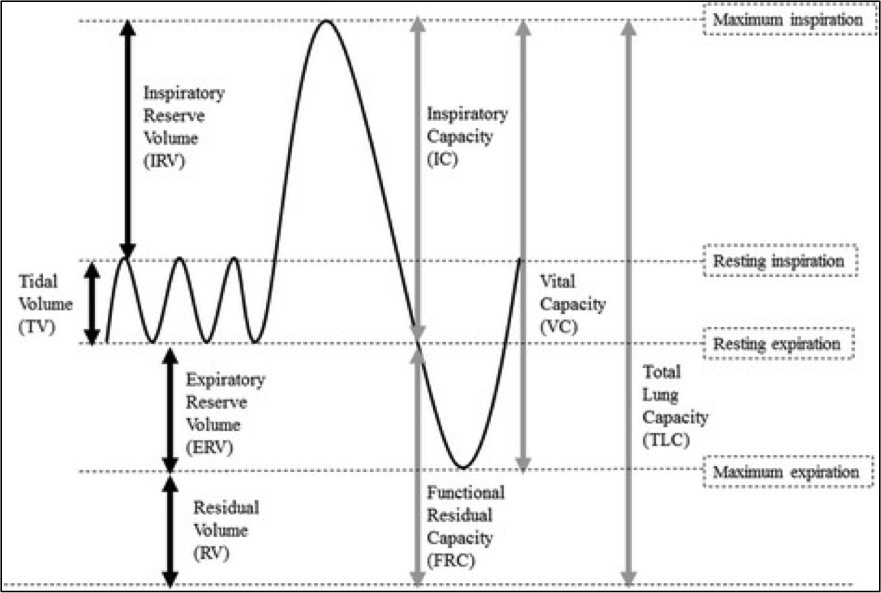
Standard Lung Volumes and Capacities. Standard lung volumes and capacities that may be affected by pulmonary disease.
Contributed by Lutfi MF. The physiological basis and clinical significance of lung volume measurements. Multidisciplinary Respiratory Medicine. 2017;12:3. (CC by 4.0; Creative Commons Attribution 4.0 International License)
References
Hallett S, Toro F, Ashurst JV. Physiology, Tidal Volume. StatPearls. 2024 Jan:(): [PubMed PMID: 29494108]
Lofrese JJ, Tupper C, Denault D, Lappin SL. Physiology, Residual Volume. StatPearls. 2024 Jan:(): [PubMed PMID: 29630222]
Ponce MC, Sankari A, Sharma S. Pulmonary Function Tests. StatPearls. 2024 Jan:(): [PubMed PMID: 29493964]
Huprikar NA, Skabelund AJ, Bedsole VG, Sjulin TJ, Karandikar AV, Aden JK, Morris MJ. Comparison of Forced and Slow Vital Capacity Maneuvers in Defining Airway Obstruction. Respiratory care. 2019 Jul:64(7):786-792. doi: 10.4187/respcare.06419. Epub 2019 Mar 19 [PubMed PMID: 30890630]
. Testing your lungs: spirometry. Breathe (Sheffield, England). 2018 Sep:14(3):257-260. doi: 10.1183/20734735.ELF143. Epub [PubMed PMID: 30186530]
Zimmermann SC, Tonga KO, Thamrin C. Dismantling airway disease with the use of new pulmonary function indices. European respiratory review : an official journal of the European Respiratory Society. 2019 Mar 31:28(151):. doi: 10.1183/16000617.0122-2018. Epub 2019 Mar 27 [PubMed PMID: 30918023]
Krishna R, Chapman K, Ullah S. Idiopathic Pulmonary Fibrosis. StatPearls. 2024 Jan:(): [PubMed PMID: 28846333]
van Gaal SC, English SW, Bourque PRJ, Zwicker JC. Pulmonary Function Testing in Elderly Patients Treated for a Myasthenia Gravis Exacerbation. The Neurohospitalist. 2019 Apr:9(2):79-84. doi: 10.1177/1941874418811249. Epub 2018 Nov 26 [PubMed PMID: 30915185]