Definition/Introduction
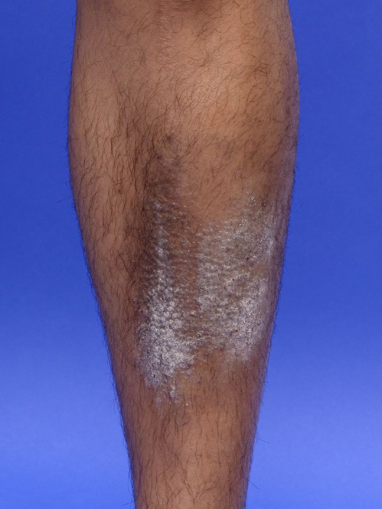
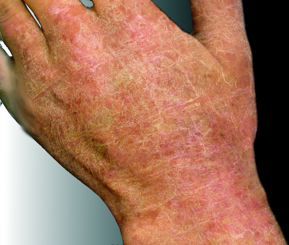
Basic skin lesions divide into primary, secondary, and special types. The term lichenification is classed as a secondary skin lesion wherein the characteristic features of skin thickening, hyperpigmentation, and exaggerated skin lines are noted. Lichenification can be further divided into primary and secondary types wherein primary lichenification signifies lichen simplex chronicus, also known as neurodermatitis circumscripta. On the other hand, secondary lichenification occurs in atopic dermatitis, infective eczematous dermatoses, psoriasis, psoriasiform dermatosis, xerosis, pityriasis rubra pilaris, porokeratosis, vegetative growths, anxiety, and obsessive-compulsive disorders.[1][2][3]
The lesions start as hyperpigmentation on a flat skin surface that is followed by the appearance of multiple small papules, termed pseudopapules, giving the lesion a pebbly appearance. The next is thickening or induration of the deep layers of the skin that does not involve the skin creases and thereby imparts the rather characteristic exaggeration of skin markings. Lesions appear as ill-defined except for certain cases of secondary lichenification.[4][5]
Owing to the inciting factor of chronic rubbing, lichenification often occurs in easily accessible sites such as the nape of the neck, wrists, hands, forearms, waist, scrotum, vulva, thighs, lower legs and dorsa of feet.[6] Certain cases are known to occur in the lower eyelids, postauricular area, axillae, and popliteal fossae.[7][8]
Diffuse types of lichenification have been described wherein involvement of the face, neck, chest, abdomen, flanks, arms, forearms, thighs and lower legs appear as ill-defined plaques ranging from hyperpigmented patches, numerous flat-topped papules, and indurated plaques may present symmetrically with extremity involvement.[9][10]
Issues of Concern
Register For Free And Read The Full Article
Search engine and full access to all medical articles
10 free questions in your specialty
Free CME/CE Activities
Free daily question in your email
Save favorite articles to your dashboard
Emails offering discounts
Learn more about a Subscription to StatPearls Point-of-Care
Issues of Concern
The presence of lichenified plaques in a healthy individual necessitates a workup for systemic disorders that present as chronic pruritus and psychological evaluation for anxiety, obsessive-compulsive disorders, or psychogenic.[11][12]
Diffuse lichenification can be a manifestation of cutaneous T-cell lymphoma and Sezary syndrome, particularly the folliculotropic type.[13][14]
Reports of chronic pruritus as a paraneoplastic phenomenon necessitates thorough screening in cases with a high index of suspicion. These include [15]:
- Hodgkin’s disease
- Lymphoma and leukemia
- Multiple myeloma
- Adenocarcinoma of the gastrointestinal tract
- Lung carcinoma
- Pancreatic carcinoma
- Insulinoma
Clinical Significance
Lichenification can be a clinical sign of underlying systemic disorders necessitating workup for psychological, malignant, metabolic, and even autoimmune disorders.
Lichenification is makes up part of the minor criteria under Hanifin and Rajka’s criteria for the diagnosis of atopic dermatitis. Other signs in close similarity include Dennie-Morgan fold, anterior neck folds, and hyperlinearity of palms and soles.[16]
The classical leathery or tree bark sign in lichen simplex chronicus represents lichenification.[6][17]
Media
(Click Image to Enlarge)
References
Voicu C, Tebeica T, Zanardelli M, Mangarov H, Lotti T, Wollina U, Lotti J, França K, Batashki A, Tchernev G. Lichen Simplex Chronicus as an Essential Part of the Dermatologic Masquerade. Open access Macedonian journal of medical sciences. 2017 Jul 25:5(4):556-557. doi: 10.3889/oamjms.2017.133. Epub 2017 Jul 24 [PubMed PMID: 28785363]
Wick MR. Psoriasiform dermatitides: A brief review. Seminars in diagnostic pathology. 2017 May:34(3):220-225. doi: 10.1053/j.semdp.2016.12.006. Epub 2016 Dec 14 [PubMed PMID: 28094165]
Granlund H, Remitz A, Kyllönen H, Lauerma AI, Reitamo S. Treatment of lichenified atopic eczema with tacrolimus ointment. Acta dermato-venereologica. 2001 Aug-Sep:81(4):314-5 [PubMed PMID: 11720192]
Level 3 (low-level) evidenceMorris M, Prurigo, Pruriginous Eczema, and Lichenification. British medical journal. 1912 Jun 29; [PubMed PMID: 20766224]
Bargout R, Malhotra A. Lower leg edema and lichenification. Elephantiasis nostras verrucosa. Postgraduate medicine. 2001 May:109(5):167-8 [PubMed PMID: 11381666]
Level 3 (low-level) evidenceCharifa A, Badri T, Harris BW. Lichen Simplex Chronicus. StatPearls. 2023 Jan:(): [PubMed PMID: 29763167]
Arseculeratne G, Altmann P, Millard PR, Todd P, Wojnarowska F. Giant lichenification of the scalp. Clinical and experimental dermatology. 2003 May:28(3):257-9 [PubMed PMID: 12780706]
Level 3 (low-level) evidenceGoldstein RK,Bastian BC,Elsner P,Burg G, Giant lichenification of the vulva with marked ulcerations. A case report. The Journal of reproductive medicine. 1991 Apr; [PubMed PMID: 2072363]
Level 3 (low-level) evidenceLavery MJ, Stull C, Kinney MO, Yosipovitch G. Nocturnal Pruritus: The Battle for a Peaceful Night's Sleep. International journal of molecular sciences. 2016 Mar 22:17(3):425. doi: 10.3390/ijms17030425. Epub 2016 Mar 22 [PubMed PMID: 27011178]
MARTINA G. [Diffuse giant lichenification]. Minerva dermatologica. 1962 Feb:37():75-7 [PubMed PMID: 14470465]
Level 3 (low-level) evidenceMorris M. A Discussion on Prurigo, Pruriginous Eczema, and Lichenification. Proceedings of the Royal Society of Medicine. 1912:5(Dermatol Sect):199-200 [PubMed PMID: 19975772]
Sequeira. A Discussion on Prurigo, Pruriginous Eczema, and Lichenification. Proceedings of the Royal Society of Medicine. 1912:5(Dermatol Sect):198-9 [PubMed PMID: 19975770]
Ehsani AH, Azizpour A, Noormohammadpoor P, Seirafi H, Farnaghi F, Kamyab-Hesari K, Sharifi M, Nasimi M. Folliculotropic Mycosis Fungoides: Clinical and Histologic Features in Five Patients. Indian journal of dermatology. 2016 Sep-Oct:61(5):554-8. doi: 10.4103/0019-5154.190124. Epub [PubMed PMID: 27688448]
Demirkesen C, Esirgen G, Engin B, Songur A, Oğuz O. The clinical features and histopathologic patterns of folliculotropic mycosis fungoides in a series of 38 cases. Journal of cutaneous pathology. 2015 Jan:42(1):22-31. doi: 10.1111/cup.12423. Epub 2014 Dec 8 [PubMed PMID: 25376535]
Level 3 (low-level) evidenceWeisshaar E, Weiss M, Mettang T, Yosipovitch G, Zylicz Z, Special Interest Group of the International Forum on the Study of Itch. Paraneoplastic itch: an expert position statement from the Special Interest Group (SIG) of the International Forum on the Study of Itch (IFSI). Acta dermato-venereologica. 2015 Mar:95(3):261-5. doi: 10.2340/00015555-1959. Epub [PubMed PMID: 25179683]
Hanifin JM, Progress in Understanding Atopic Dermatitis. The Journal of investigative dermatology. 2018 Dec; [PubMed PMID: 30466540]
Level 3 (low-level) evidenceRajalakshmi R, Thappa DM, Jaisankar TJ, Nath AK. Lichen simplex chronicus of anogenital region: a clinico-etiological study. Indian journal of dermatology, venereology and leprology. 2011 Jan-Feb:77(1):28-36. doi: 10.4103/0378-6323.74970. Epub [PubMed PMID: 21220876]
Level 2 (mid-level) evidence
