Introduction
A herald patch is a distinct skin manifestation characterized by a single, erythematous, round to oval scaly patch or plaque. The patch has a depressed center and raised border and measures 2 to 10 cm (see Image. Herald Patch). The lesion typically exhibits a predilection for the neck, chest, and back. As the lesion evolves, it tends to clear centrally, leaving the free edge of the scale. This reveals a unique "collarette" of scale, which is a common presenting sign of pityriasis rosea—a prevalent and self-limiting papulosquamous disorder. This clinical presentation resembles the edge of a cigarette paper directed inward toward the center.
The sign is called the "herald" patch or "mother" patch because it often appears a few days to 2 weeks before the complete eruption of pityriasis rosea.[1][2][3][4] In 10% to 50% of the cases of pityriasis rosea, the herald patch may be absent, especially in drug-induced cases. On the other hand, the herald patch may appear in multiple locations and atypical sites, such as the soles or scalp. In some cases, the skin patch is the sole manifestation of the disease, not followed by a secondary rash.
Etiology
Register For Free And Read The Full Article
 Search engine and full access to all medical articles
- 10 free questions in your specialty
- Free CME/CE Activities
- Free daily question in your email
- Save favorite articles to your dashboard
- Emails offering discounts
Learn more about a Subscription to StatPearls Point-of-Care
Etiology
The herald patch and pityriasis rosea are thought to be caused by infection with human herpesvirus 6 (HHV-6) and human herpesvirus 7 (HHV-7), but this is debatable. Evidence supporting an infectious etiology includes outbreaks occurring in clusters, the prodromal symptoms in most patients before the herald patch or eruption of pityriasis rosea occurs, and the rare instance of recurrence that suggests immunity. Human herpesvirus 8 (HHV-8), the 2009 to 2010 pandemic H1N1 influenza A virus, and severe acute respiratory syndrome coronavirus 2 (SARS-CoV-2) have been reported in association with pityriasis rosea.[5][6][7][8]
In one retrospective cohort study, pityriasis rosea only recurred in 3.7% of patients, but none of these recurrences manifested with a herald patch. Interactions between Langerhans cells, unknown components of the epidermis, and dermal dendritic cells have also been postulated, but whether these interactions are due to a viral etiology is still uncertain.[9][10][11] Pityriasis rosea has also been linked with vaccinations for smallpox, tuberculosis, influenza, papillomavirus, polio, tetanus, diphtheria, pneumococcal, diphtheria-pertussis-tetanus, hepatitis B, and yellow fever [12] and more recently with the COVID-19 vaccines.[13][14] Pityriasis rosea-like eruptions can also occur in association with many drugs, such as acetylsalicylic acid, barbiturates, bismuth, captopril, clonidine, gold, imatinib, isotretinoin, ketotifen, levamisole, metronidazole, omeprazole, D-penicillamine, and terbinafine.[15]
Epidemiology
The herald patch is seen in approximately 80% of the cases of pityriasis rosea. The incidence of pityriasis rosea peaks between 20 and 29, but it may be seen in any age group. Pityriasis rosea occurs slightly more in females than in males.[16] The disease is more common in the spring and the fall in temperate climate zones. No racial predominance is reported. Nonetheless, intensely pigmented Africans tend to have more widespread disease, with lesions darker than the surrounding skin, unlike the rose color seen in other patients with fair complexion.
Pathophysiology
The pathophysiology of pityriasis rosea, and thus the herald patch, is poorly understood. As stated, an association with HHV-6 and HHV-7 is recognized in the plasma and lesional skin of patients with pityriasis rosea, but other studies have revealed conflicting evidence. The inflammatory populations seen in lesions of pityriasis rosea and the herald patch are predominantly T cells with a lack of B cells and natural killer cells. Natural killer cells are cytotoxic to cells infected with viruses, and their presence is suspected if it is solely due to a viral etiology. Interactions between the Langerhans cells and unknown components of the epidermis have been implicated in the pathophysiology, but this is not fully understood.
Histopathology
The immunohistochemical and histopathological findings in the herald patch and fully developed lesions of pityriasis rosea are similar. Histological findings of pityriasis rosea include focal or confluent parakeratosis, epidermal hyperplasia, spongiosis, exocytosis of lymphocytes, and extravasation of erythrocytes along with a moderately dense perivascular lymphocytic infiltrate in the superficial dermis. Due to its chronic nature, the herald patch has similar features but a deeper infiltrate and more acanthosis.
Some other variations have been observed, such as dyskeratotic cells in the epidermis, multinuclear giant cells, and focal acantholytic dysfunction. These features may closely resemble erythema annulare centrifugum, guttate psoriasis, superficial gyrate erythema, and small plaque parapsoriasis. Immunohistochemical analysis usually reveals an increased CD4+ to a CD8+ ratio of T lymphocytes, and increased numbers of Langerhans cells have been observed in specimens.
History and Physical
The herald patch is a round to oval erythematous patch or plaque with central clearing and peripheral scale trailing behind the margins of the erythema. The herald patch typically measures 2 to 10 cm. However, rare reports of very large herald patches have been described, with one case nearly covering the entire trunk of an 18-month-old female child.[17] Most herald patches appear on the trunk, neck, or proximal extremities, although in rare cases, they have appeared on the face, genitalia, scalp, and acral surfaces. Most lesions are asymptomatic, but some may be pruritic.
Most patients experience prodromal symptoms, including a headache, fever, arthralgias, and malaise before or around the time the herald patch appears. A few days to 2 weeks after the appearance of the herald patch, patients develop a more diffuse eruption of pityriasis rosea. This is accompanied by smaller lesions resembling the herald patch along skin cleavage lines in a "Christmas tree" or "fir tree" distribution, with truncal predominance. The lesions are sometimes atypical in children; they may be follicular-papular, pustular, vesicular, urticarial, or purpuric.[18] The eruption spreads from the top down over a few days. The eruption fades in 4 to 6 weeks, leaving few residual changes, such as postinflammatory dyspigmentation. Oropharyngeal abnormalities have been reported in patients with pityriasis rosea in the form of petechial, macular, and papular eruptions.[19] A proposed classification divides pityriasis rosea into classic, relapsing, persistent, pediatric, pityriasis rosea in pregnancy, and pityriasis rosea–like eruptions.[20]
Evaluation
A definitive diagnosis of the herald patch can be difficult before the classic eruption of pityriasis rosea. Typically, no laboratory abnormalities in pityriasis rosea are expected. Due to the morphological similarities with tinea corporis, a potassium hydroxide (KOH) examination of scales for dermatophyte hyphae may be necessary to distinguish these conditions. If the potassium hydroxide examination is equivocal, a biopsy may be performed, which may aid in differentiating the herald patch from tinea, erythema annular centrifugum, or nummular eczema. When the full eruption of pityriasis rosea finally presents, the palms and soles should be checked for involvement, as the eruption can mimic secondary syphilis. If secondary syphilis cannot be ruled out, nontreponemal tests such as the rapid plasma reagin test should be performed with appropriate confirmatory tests.[21][22][23][24]
Treatment / Management
Because the herald patch is a feature of pityriasis rosea, the same treatment is advised for both. The eruption of pityriasis rosea is a benign, self-limited papulosquamous process. No treatment is necessary, and the eruption of both the herald patch and pityriasis rosea should resolve within 8 weeks. Patients typically seek treatment for pruritis that occasionally is associated with pityriasis rosea, or because the appearance of the rash perturbs them. Most cases of pityriasis rosea are asymptomatic or minimally pruritic, but approximately 25% of patients experience severe pruritis.
If the lesions are symptomatic or disturbing, the following guidance is beneficial:
- Patients and their caregivers should be reassured that pityriasis rosea typically resolves within 2 to 3 months with low risk for transmission, and recurrence is uncommon. Patients should also be instructed to avoid scratching and contact with irritants, such as harsh soaps, synthetic fabrics, fragrances, hot water, tight clothing, and sweating.
- Topical steroids in the medium potency range should be applied to the pruritic areas 2 to 3 times per day for 2 to 3 weeks, under medical supervision, to avoid corticosteroid-induced skin atrophy that could result from long-term use.
- Topical antipruritic lotions that contain pramoxine, menthol, calamine lotion, or zinc oxide may help to reduce pruritus.[25]
- Oral antihistamines can reduce the irritation and help the patients sleep better at night.
- Oral steroids may also improve the pruritus of pityriasis rosea; however, routine use is not recommended due to limited data on their efficacy and concerns of relapse after treatment.[26] (A1)
- The postulated link between pityriasis rosea and human herpes viruses led to the investigation of oral acyclovir in patients with pityriasis rosea. However, high-quality studies are still lacking, and the rationale for its benefit remains unclear.[27]
- Ultraviolet B phototherapy and a 2-week course of oral erythromycin have been shown to shorten the duration of the eruption.[28][29][28] (A1)
- UVA1 phototherapy is associated with reduced disease severity and improvement in pruritus.[30]
Differential Diagnosis
The differential diagnosis of the herald patch differs from pityriasis rosea because the former is a single lesion while the latter is an eruption. The differential diagnosis for the herald patch includes tinea corporis, nummular eczema, and erythema annulare centrifugum. A helpful distinguishing feature among most of these conditions is the location of the scale within the erythema. The herald patch and erythema annulare centrifugum often exhibit a trailing scale. This is seen as a collarette of scale inside the borders of the erythema. The edge of the scale in tinea appears to lead the erythema in an annular fashion. Nummular eczema typically has scale throughout the lesion without central clearing.
When herald patch evolves into the complete picture of pityriasis rosea, the differential diagnosis should include secondary syphilis, guttate psoriasis, tinea versicolor, and pityriasis lichenoides chronica. Lyme disease, HIV, seroconversion illness, and drug eruptions should also be considered in the differential diagnosis of pityriasis rosea. Testing for HIV should be performed in patients with risk factors for or symptoms suggestive of HIV infection.
Prognosis
The eruption of the herald patch and pityriasis rosea should resolve within 8 weeks. Reports of a variant of pityriasis rosea known as persistent pityriasis rosea have been described, with some lasting longer than 12 weeks. The herald patch is seen in most cases, and a higher association with systemic symptoms, oral lesions, and increased HHV-6 and HHV-7 viral loads was found in association. Relapse after a resolution is uncommon (approximately 2%).[33]
Complications
Postinflammatory hyperpigmentation is a common sequela in individuals with darkly pigmented skin and often takes several months or longer to resolve. Research on the impact of pityriasis rosea on pregnancy is limited and conflicting. An analysis of a case series of women who developed the disease during pregnancy suggested that pityriasis rosea increases the risk of spontaneous abortion.[34] However, the frequency of spontaneous abortion is lower in other series and comparable with rates in patients without the disease.[35][36]
An analysis of pooled data from patients in case reports and case series suggested a greater likelihood for unfavorable pregnancy outcomes, such as preterm delivery, low birth weight, or spontaneous abortion, among pregnant individuals with extensive or prolonged course of the disease, onset of eruption earlier in pregnancy, or associated extracutaneous symptoms.[35] However, the timing of the onset of pityriasis rosea did not seem to be a relevant factor for pregnancy complications in another case series.[36] Further studies are necessary to clarify the impact of pityriasis rosea on pregnancy.
Deterrence and Patient Education
Patients affected by the herald patch and its subsequent secondary rash of pityriasis rosea should be educated about the self-limiting and noninfectious nature of the disease. Treatment is mainly supportive to relieve the symptoms of itching. Patients should avoid scratching the lesions and contact with irritants.
Pearls and Other Issues
Herald patch is the first to present a lesion of pityriasis rosea. Although the presentation is unique to pityriasis rosea, it can resemble tinea corporis, discoid eczema, and erythema annulare centrifugum. Within 2 weeks, the distinctive secondary rash of pityriasis rosea develops following the cleavage lines of the skin, forming the "Christmas tree" pattern. This persists for another 2 weeks and resolves over another 2 weeks without needing treatment. Nonetheless, some lesions may persist for 3 to 4 months.
Enhancing Healthcare Team Outcomes
Pityriasis rosea is a commonly encountered skin condition in clinical practice, managed by primary care clinicians, nurse practitioners, dermatologists, and emergency department clinicians. Recognizing the herald patch is crucial, as at least 25% of patients experience severe itching, which, if left untreated, can significantly impact their quality of life. These patients require follow-up as recurrences are possible.[37]
Media
(Click Image to Enlarge)
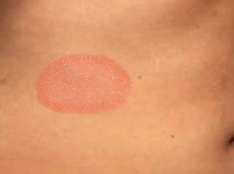
Herald Patch. A herald patch presents as a circular or oval-shaped erythematous patch with central clearing and trailing peripheral scales measuring 2 to 10 cm.
Contributed by S Bhimji, MD
References
Singh M, Pawar M, Chuh A, Zawar V. Pityriasis rosea: elucidation of environmental factors in modulated autoagressive etiology and dengue virus infection. Acta dermatovenerologica Alpina, Pannonica, et Adriatica. 2019 Mar:28(1):15-20 [PubMed PMID: 30901064]
Yüksel M. Pityriasis Rosea Recurrence is Much Higher than Previously Known: A Prospective Study. Acta dermato-venereologica. 2019 Jun 1:99(7):664-667. doi: 10.2340/00015555-3169. Epub [PubMed PMID: 30848285]
Chhabra N, Prabha N, Kulkarni S, Ganguly S. Pityriasis Rosea: Clinical Profile from Central India. Indian dermatology online journal. 2018 Nov-Dec:9(6):414-417. doi: 10.4103/idoj.IDOJ_12_18. Epub [PubMed PMID: 30505781]
Ivars M, Martin-Santiago A, Baselga E, Guibaud L, López-Gutiérrez JC. Fern-shaped patch as a hallmark of blue rubber bleb nevus syndrome in neonatal venous malformations. European journal of pediatrics. 2018 Sep:177(9):1395-1398. doi: 10.1007/s00431-018-3126-x. Epub 2018 Mar 8 [PubMed PMID: 29520504]
Veraldi S, Spigariolo CB. Pityriasis rosea and COVID-19. Journal of medical virology. 2021 Jul:93(7):4068. doi: 10.1002/jmv.26679. Epub 2020 Dec 1 [PubMed PMID: 33205836]
Martora F, Picone V, Fornaro L, Fabbrocini G, Marasca C. Can COVID-19 cause atypical forms of pityriasis rosea refractory to conventional therapies? Journal of medical virology. 2022 Apr:94(4):1292-1293. doi: 10.1002/jmv.27535. Epub 2021 Dec 31 [PubMed PMID: 34931329]
Prantsidis A, Rigopoulos D, Papatheodorou G, Menounos P, Gregoriou S, Alexiou-Mousatou I, Katsambas A. Detection of human herpesvirus 8 in the skin of patients with pityriasis rosea. Acta dermato-venereologica. 2009 Nov:89(6):604-6. doi: 10.2340/00015555-0703. Epub [PubMed PMID: 19997691]
Level 2 (mid-level) evidenceKwon NH, Kim JE, Cho BK, Park HJ. A novel influenza a (H1N1) virus as a possible cause of pityriasis rosea? Journal of the European Academy of Dermatology and Venereology : JEADV. 2011 Mar:25(3):368-9. doi: 10.1111/j.1468-3083.2010.03725.x. Epub [PubMed PMID: 20561127]
Level 3 (low-level) evidenceVillalon-Gomez JM. Pityriasis Rosea: Diagnosis and Treatment. American family physician. 2018 Jan 1:97(1):38-44 [PubMed PMID: 29365241]
Litchman G, Nair PA, Le JK. Pityriasis Rosea. StatPearls. 2024 Jan:(): [PubMed PMID: 28846360]
Gupta N, Levitt JO. Unique clinical presentations of pityriasis rosea: aphthous ulcers, vesicles and inverse distribution of lesions. Dermatology online journal. 2017 Feb 15:23(2):. pii: 13030/qt3mk4z6w0. Epub 2017 Feb 15 [PubMed PMID: 28329497]
Drago F, Ciccarese G, Javor S, Parodi A. Vaccine-induced pityriasis rosea and pityriasis rosea-like eruptions: a review of the literature. Journal of the European Academy of Dermatology and Venereology : JEADV. 2016 Mar:30(3):544-5. doi: 10.1111/jdv.12942. Epub 2014 Dec 29 [PubMed PMID: 25545307]
Level 3 (low-level) evidenceMartora F, Fabbrocini G, Marasca C. Pityriasis rosea after Moderna mRNA-1273 vaccine: A case series. Dermatologic therapy. 2022 Feb:35(2):e15225. doi: 10.1111/dth.15225. Epub 2021 Dec 1 [PubMed PMID: 34816549]
Level 2 (mid-level) evidenceDrago F, Broccolo F, Ciccarese G. Pityriasis rosea, pityriasis rosea-like eruptions, and herpes zoster in the setting of COVID-19 and COVID-19 vaccination. Clinics in dermatology. 2022 Sep-Oct:40(5):586-590. doi: 10.1016/j.clindermatol.2022.01.002. Epub 2022 Jan 31 [PubMed PMID: 35093476]
González LM, Allen R, Janniger CK, Schwartz RA. Pityriasis rosea: an important papulosquamous disorder. International journal of dermatology. 2005 Sep:44(9):757-64 [PubMed PMID: 16135147]
Chuang TY, Ilstrup DM, Perry HO, Kurland LT. Pityriasis rosea in Rochester, Minnesota, 1969 to 1978. Journal of the American Academy of Dermatology. 1982 Jul:7(1):80-9 [PubMed PMID: 6980904]
Level 2 (mid-level) evidenceChuh A. A Herald Patch Almost Encircling the Trunk-Extreme Pityriasis Rosea Gigantea in a Young Child. Pediatric dermatology. 2016 Sep:33(5):e286-7. doi: 10.1111/pde.12923. Epub 2016 Jul 11 [PubMed PMID: 27396667]
Chuh AA. Quality of life in children with pityriasis rosea: a prospective case control study. Pediatric dermatology. 2003 Nov-Dec:20(6):474-8 [PubMed PMID: 14651563]
Level 2 (mid-level) evidenceCiccarese G, Broccolo F, Rebora A, Parodi A, Drago F. Oropharyngeal lesions in pityriasis rosea. Journal of the American Academy of Dermatology. 2017 Nov:77(5):833-837.e4. doi: 10.1016/j.jaad.2017.06.033. Epub 2017 Jul 18 [PubMed PMID: 28728872]
Drago F, Ciccarese G, Rebora A, Broccolo F, Parodi A. Pityriasis Rosea: A Comprehensive Classification. Dermatology (Basel, Switzerland). 2016:232(4):431-7. doi: 10.1159/000445375. Epub 2016 Apr 21 [PubMed PMID: 27096928]
Çölgeçen E, Kader Ç, Ulaş Y, Öztürk P, Küçük Ö, Balcı M. Pityriasis rosea: a natural history of pediatric cases in theCentral Anatolia Region of Turkey. Turkish journal of medical sciences. 2016 Dec 20:46(6):1740-1742. doi: 10.3906/sag-1507-30. Epub 2016 Dec 20 [PubMed PMID: 28081320]
Level 3 (low-level) evidenceChuh A, Zawar V, Sciallis GF, Lee A. The diagnostic criteria of pityriasis rosea and Gianotti-Crosti syndrome - a protocol to establish diagnostic criteria of skin diseases. The journal of the Royal College of Physicians of Edinburgh. 2015:45(3):218-25. doi: 10.4997/JRCPE.2015.310. Epub [PubMed PMID: 26517103]
Allmon A, Deane K, Martin KL. Common Skin Rashes in Children. American family physician. 2015 Aug 1:92(3):211-6 [PubMed PMID: 26280141]
Ganguly S. A clinicoepidemiological study of pityriasis rosea in South India. Skinmed. 2013 May-Jun:11(3):141-6 [PubMed PMID: 23930352]
Level 2 (mid-level) evidenceChuh A, Zawar V, Sciallis G, Kempf W. A position statement on the management of patients with pityriasis rosea. Journal of the European Academy of Dermatology and Venereology : JEADV. 2016 Oct:30(10):1670-1681. doi: 10.1111/jdv.13826. Epub 2016 Jul 13 [PubMed PMID: 27406919]
Sonthalia S, Kumar A, Zawar V, Priya A, Yadav P, Srivastava S, Gupta A. Double-blind randomized placebo-controlled trial to evaluate the efficacy and safety of short-course low-dose oral prednisolone in pityriasis rosea. The Journal of dermatological treatment. 2018 Sep:29(6):617-622. doi: 10.1080/09546634.2018.1430302. Epub 2018 Feb 1 [PubMed PMID: 29363373]
Level 1 (high-level) evidenceDe Clercq E, Naesens L, De Bolle L, Schols D, Zhang Y, Neyts J. Antiviral agents active against human herpesviruses HHV-6, HHV-7 and HHV-8. Reviews in medical virology. 2001 Nov-Dec:11(6):381-95 [PubMed PMID: 11747000]
Sharma PK, Yadav TP, Gautam RK, Taneja N, Satyanarayana L. Erythromycin in pityriasis rosea: A double-blind, placebo-controlled clinical trial. Journal of the American Academy of Dermatology. 2000 Feb:42(2 Pt 1):241-4 [PubMed PMID: 10642679]
Level 1 (high-level) evidenceValkova S, Trashlieva M, Christova P. UVB phototherapy for Pityriasis rosea. Journal of the European Academy of Dermatology and Venereology : JEADV. 2004 Jan:18(1):111-2 [PubMed PMID: 14678553]
Level 3 (low-level) evidenceLim SH, Kim SM, Oh BH, Ko JH, Lee YW, Choe YB, Ahn KJ. Low-dose Ultraviolet A1 Phototherapy for Treating Pityriasis Rosea. Annals of dermatology. 2009 Aug:21(3):230-6. doi: 10.5021/ad.2009.21.3.230. Epub 2009 Aug 31 [PubMed PMID: 20523795]
Amer A, Fischer H. Azithromycin does not cure pityriasis rosea. Pediatrics. 2006 May:117(5):1702-5 [PubMed PMID: 16651327]
Level 1 (high-level) evidenceAhmed N, Iftikhar N, Bashir U, Rizvi SD, Sheikh ZI, Manzur A. Efficacy of clarithromycin in pityriasis rosea. Journal of the College of Physicians and Surgeons--Pakistan : JCPSP. 2014 Nov:24(11):802-5 [PubMed PMID: 25404436]
Level 1 (high-level) evidenceDrago F, Broccolo F, Rebora A. Pityriasis rosea: an update with a critical appraisal of its possible herpesviral etiology. Journal of the American Academy of Dermatology. 2009 Aug:61(2):303-18. doi: 10.1016/j.jaad.2008.07.045. Epub [PubMed PMID: 19615540]
Drago F, Broccolo F, Javor S, Drago F, Rebora A, Parodi A. Evidence of human herpesvirus-6 and -7 reactivation in miscarrying women with pityriasis rosea. Journal of the American Academy of Dermatology. 2014 Jul:71(1):198-9. doi: 10.1016/j.jaad.2014.02.023. Epub [PubMed PMID: 24947696]
Level 3 (low-level) evidenceWenger-Oehn L, Graier T, Ambros-Rudolph C, Müllegger R, Bittighofer C, Wolf P, Hofer A. Pityriasis rosea in pregnancy: A case series and literature review. Journal der Deutschen Dermatologischen Gesellschaft = Journal of the German Society of Dermatology : JDDG. 2022 Jul:20(7):953-959. doi: 10.1111/ddg.14763. Epub 2022 May 26 [PubMed PMID: 35616213]
Level 2 (mid-level) evidenceStashower J, Bruch K, Mosby A, Boddie PP, Varghese JA, Rangel SM, Brodell RT, Zheng L, Flowers RH. Pregnancy complications associated with pityriasis rosea: A multicenter retrospective study. Journal of the American Academy of Dermatology. 2021 Dec:85(6):1648-1649. doi: 10.1016/j.jaad.2020.12.063. Epub 2021 Jan 8 [PubMed PMID: 33422632]
Level 2 (mid-level) evidenceChuah SY, Chia HY, Tan HH. Recurrent and persistent pityriasis rosea: an atypical case presentation. Singapore medical journal. 2014 Jan:55(1):e4-6 [PubMed PMID: 24452984]
Level 3 (low-level) evidence