Introduction
Ankle arthrocentesis is a procedure that determines the underlying etiology of monoarthropathy of the ankle. The underlying etiology may be unclear for monoarthropathy of a joint, as the clinical characteristics of monoarthropathy and the differential diagnosis may overlap. The values from synovial fluid analysis following arthrocentesis can help clinicians distinguish between a non-urgent inflammatory process requiring anti-inflammatory and analgesic therapy and an urgent infectious process requiring antibiotics and surgical intervention.[1][2]
Many etiologies of monoarthropathy may be evident after obtaining patient history (eg, hemarthrosis from trauma); therefore, arthrocentesis is reserved for cases with an unclear cause. Obtaining objective data for diagnosis is paramount for adult patients, as the commonly used Kocher criteria for pediatric septic arthritis do not apply to adults. In rare cases, septic and crystal-induced arthritis may occur concurrently, and arthrocentesis is instrumental. Accessing the joint can drain a painful effusion or ensure local injection of medications to minimize pain and inflammation.[3][4] Improved ultrasound imaging introduces alternatives to arthrocentesis compared to traditional landmark-guided methods. Ultrasound increases the clinician's confidence in performing the procedure, improving accuracy. Additional research is needed to determine if ultrasound facilitates superior patient outcomes.[5][6][7]
Anatomy and Physiology
Register For Free And Read The Full Article
 Search engine and full access to all medical articles
- 10 free questions in your specialty
- Free CME/CE Activities
- Free daily question in your email
- Save favorite articles to your dashboard
- Emails offering discounts
Learn more about a Subscription to StatPearls Point-of-Care
Anatomy and Physiology
The 2 joint spaces of the ankle routinely involved in arthrocentesis are the subtalar and tibiotalar joints. The subtalar joint is formed by the talus' inferior surface and the calcaneus's superior surface. The posterior talocalcaneal, medial talocalcaneal, and lateral talocalcaneal ligaments support the joint capsule. Access to subtalar joint effusions involves the lateral malleolus and sinus tarsus anatomical structures.[1][8]
The 2 long bones of the lower leg and a tarsal bone form the tibiotalar joint. The superior surface of the talus forms the tibiotalar joint. The mortise is formed by the distal end of the tibia and fibula. The joint capsule is supported by 3 groups of ligaments: the tibiofibular syndesmosis, medial or deltoid collateral ligaments, and lateral collateral ligaments.[9] Access to tibiotalar joint effusions involves the anatomical structures of the medial malleolus, extensor hallucis longus tendon, and tibialis anterior tendon.[1][2][5]
Indications
The following are indications for arthrocentesis:
Diagnostic Arthrocentesis
- Septic or crystal-induced arthritis
- Arthritis with synovial effusion of unknown etiology
- Evaluation of septic arthritis response to therapy [10][11][12]
Therapeutic Arthrocentesis
Contraindications
Overlying skin or soft tissue infection is an absolute contraindication to ankle arthrocentesis. Relative contraindications include septic arthritis (suspected or proven), multiple corticosteroid injections, intraarticular fracture, juxta-articular osteopenia, joint instability, and inability to rest post-procedure.[1][2]
Equipment
The required equipment for ankle arthrocentesis includes:
- Sterile gloves
- Betadine or chlorhexidine prep
- Needle
- The most common size is 20-gauge
- Larger needles may be considered for thick synovial fluid
- Smaller needles may be considered for individuals with small joints
- Syringes
- 22-gauge (or smaller), short-length
- 10 mL
- 1 mL
- Lidocaine (1%)
- Tubes
- Aerobic and anaerobic sterile culture
- Ethylenediaminetetraacetic acid or heparinized
- Fluoride
- Clear glass
- Gauze
- Adhesive bandages
- Optional materials include:
- Alcohol wipe or swab
- Surgical marker
- 5 MHz linear transducer (or equivalent) with sterile probe cover
- Corticosteroids
Personnel
A skilled clinician may reasonably perform this procedure; assistance with the ultrasound or collection tubes reduces difficulty and increases the chances of success. Assisting clinicians are typically medical assistants or nurses. An assisting ultrasound technician can improve the reliability of probe use.
Preparation
Informed consent should be obtained after discussing potential risks. The patient should be in a supine or reclined position to facilitate easy access to the anterior region of the ankle and reduce any weight bearing on the ankle. This positioning allows maximum joint space during the procedure. The cutaneous region over the desired joint should be adequately cleaned using an alcohol wipe, swab, or the above preparatory options. Cutaneous analgesia can be obtained with the use of the 1% lidocaine and the 1 mL syringe.
Technique or Treatment
Subtalar Joint Without Ultrasound Guidance
- The foot is positioned so a view of the lateral aspect of the ankle is obtained (see Image. Positioning and Location to Access Subtalar Ankle Joint [Lateral View]). With the foot in a perpendicular position relative to the leg, palpate inferior and anterior to the distal tip of the lateral malleolus. When the depression is palpated in this region and associated with sinus tarsi, the area is marked. Cleaning the area and cutaneous analgesia is recommended.
- The 20-gauge needle is attached to the end of an empty 10 mL syringe, and the joint is entered by directing the needle horizontally across the ankle while remaining in line with the lateral malleolus (see Image. Positioning and Location to Access Subtalar Ankle Joint).
- Keeping negative pressure on the syringe plunger during this process and feeling for the subtle pop when penetrating the synovial membrane will alert the treating clinician when the joint has been accessed. The presence of synovial fluid in the syringe indicates the joint is accessible.
- Draining of effusion or injection of medication can commence. Due to the limited size of the ankle joints, switching syringes to continue removing effusion after the syringe is full is rarely required.
- Pressure with gauze is applied to the skin after withdrawing the needle, which an adhesive bandage or dressing can follow.[1]
Subtalar Joint With Ultrasound Guidance
- The foot is positioned so a view of the lateral aspect of the ankle can be obtained. With the foot in a perpendicular position relative to the leg, palpate anterior to the distal tip of the lateral malleolus.
- The ultrasound transducer is then placed on a short axis to the foot just anterior to the lateral malleolus directly over the sinus tarsi. The image on the screen should show the echogenic talus on one side and the echogenic calcaneus on the other, with the sinus tarsi in between. Cleaning the area and the region's cutaneous analgesia is recommended.
- Next, with the 20-gauge needle attached to the end of an empty 10 mL syringe, the joint is entered by directing the needle posteromedial towards the sinus tarsi, keeping the needle horizontally across the ankle in line with the lateral malleolus (see Image. Ultrasound With Tibial Landmarks).[13][14]
- The remainder of the procedure is the same as detailed above for the subtalar joint without ultrasound guidance.
Tibiotalar Joint Without Ultrasound Guidance
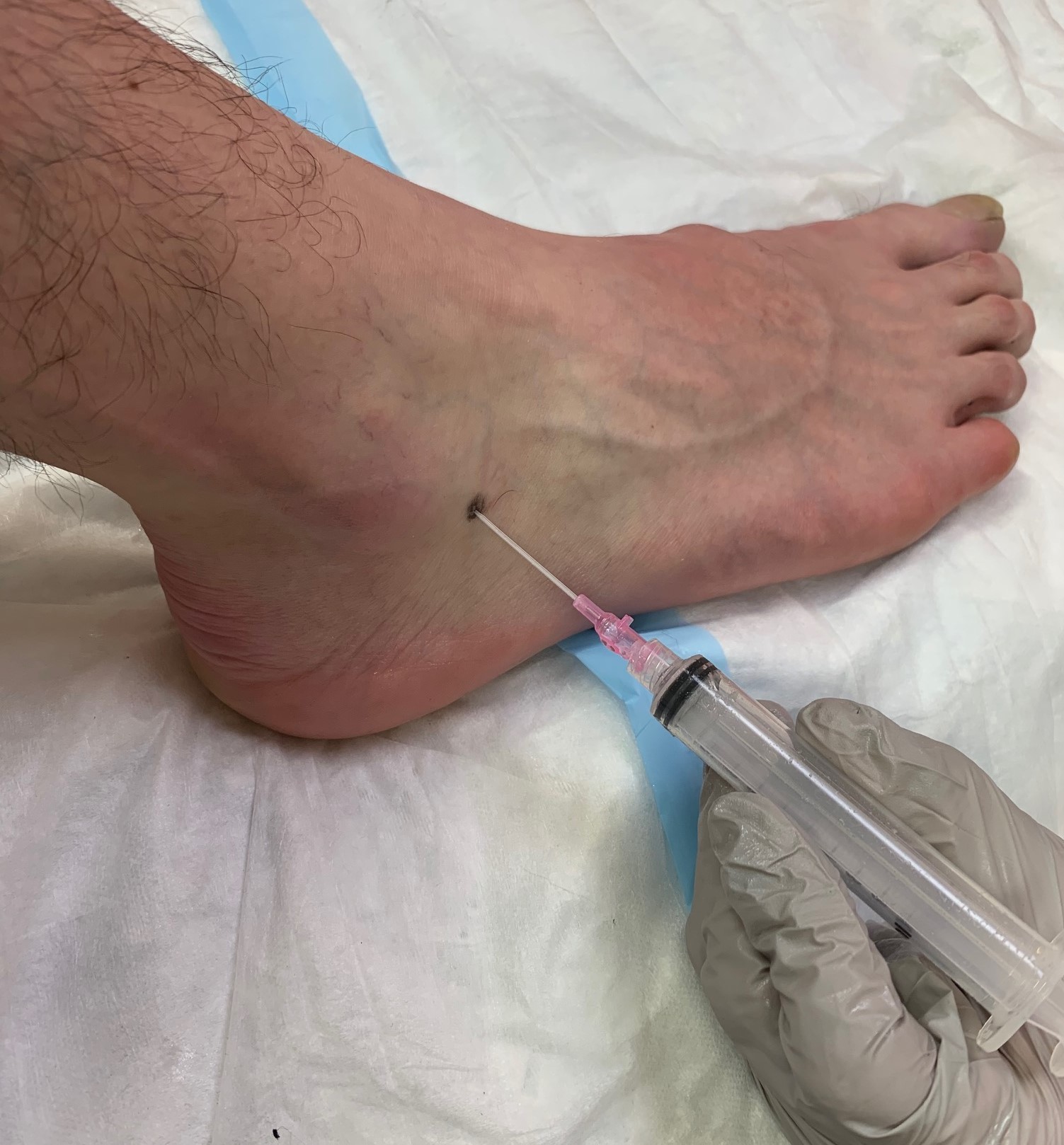
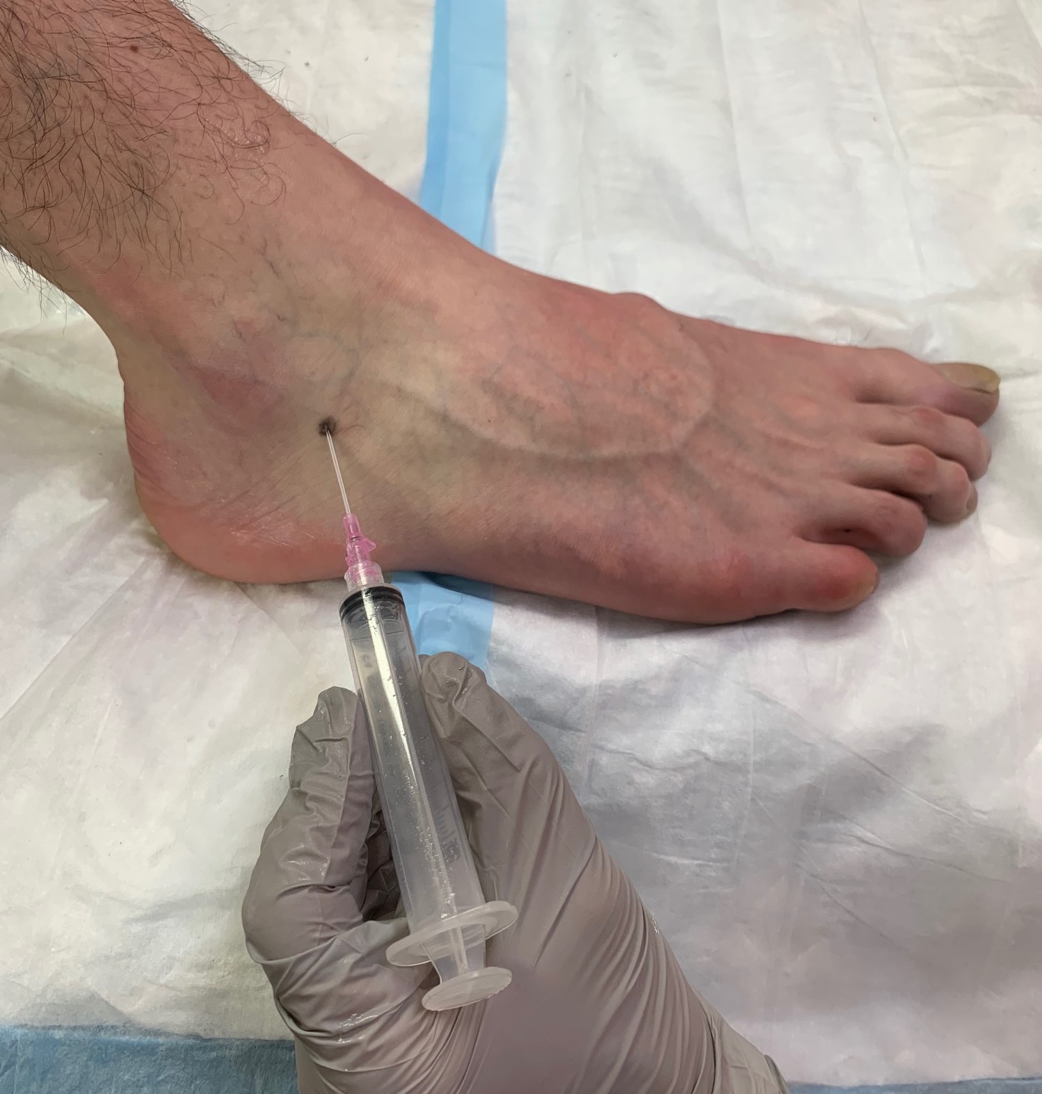
- The foot is positioned so a view of the anteromedial aspect of the ankle is obtained. With the foot in a perpendicular position relative to the leg, palpate approximately 1 cm anterior to the distal tip of the medial malleolus. Keeping posterior and medial to the extensor hallucis longus and tibialis anterior tendons (easier to palpate by asking the patient to dorsiflex their great toe), a marking is placed between the palpated medial malleolus and tendons. Cleaning the area and cutaneous analgesia of the region is recommended.
- Next, with the 20-gauge needle attached to the end of an empty 10 mL syringe, the joint is entered by directing the needle in a slightly posterolateral direction across the ankle while keeping the bevel in relatively parallel relation with the dorsum of the foot, the horizontal plane of the tibiotalar joint.
- The remainder of the procedure is the same as detailed above for the subtalar joint without ultrasound guidance.[2]
Tibiotalar Joint With Ultrasound Guidance
- The placement of the ultrasound transducer should be longitudinal and just lateral to the extensor hallucis longus and tibialis anterior tendons. Setting the ultrasound depth to approximately 4 cm should provide a view of the anterior surface of the tibia on one side and the curving dome of the talus on the other side. The desired tibiotalar joint resides between these 2 bony landmarks.
- The entrance of the syringe into the joint is performed similarly to without the ultrasound. However, the syringe's location is more precise when the ultrasound guides how proximal or distal the ankle enters the skin. Fanning the ultrasound transducer will allow the operator to visualize the entrance of the needle into the joint space.
- The remainder of the procedure is the same as detailed for the subtalar joint without ultrasound guidance.[5]
Disposition of Synovial Fluid
- Sterile tube: Send for culture for aerobic bacteria, anaerobic bacteria, and fungi
- EDTA or heparinized tube: Send for white blood cell count with differential
- Fluoride tube: Send for glucose level
- Clear glass tube: for visual inspection and other indicated studies
- Slides: Order Gram stains and polarized light microscopy for crystal examination
Complications
Complications are as follows:
- Iatrogenic joint infection (1 to 2 per 25,000 arthrocentesis)
- Soft tissue and surrounding structure damage (tendon, nerves, and blood vessels)
- Inability to successfully obtain access to the desired joint (ensure adequate analgesia). The use of ultrasound is recommended.
- Inadvertent extra-synovial injection (soft-tissue or intravascular) if an injection of medication is used with the procedure.[1][2]
Clinical Significance
Because many etiologies of monoarthropathy have overlapping features, ankle arthrocentesis may be necessary for evaluation and treatment. Using ultrasound-guided versus landmark-guided techniques may enhance the success and confidence of the clinician performing the procedure.[15]
Enhancing Healthcare Team Outcomes
Ankle arthrocentesis requires an interprofessional team effort involving several clinician specialties (eg, emergency medicine, hospitalist, orthopedist), laboratory personnel, and nursing staff. These individuals are essential in obtaining, maintaining, and evaluating the synovial samples to make the diagnosis. As with any procedure that violates the skin and synovial capsule, the risk of seeding infection is always present, and evaluating the overlying cutaneous skin for the presence of skin and soft tissue infections is imperative. Meticulous records of the procedure and administered medications are requisite. The treating clinician should collaborate with nursing staff to obtain the proper supplies, record the steps performed, and transport the obtained samples to the laboratory for analysis.
Clinicians should be cognizant of contraindications to administering medications commonly used with this procedure (eg, corticosteroids not routinely recommended for use in patients with uncontrolled diabetes). A system for promptly reporting critical, abnormal laboratory results by pathology is essential. Prompt consultation with surgical specialists for any diagnosis necessitating urgent or emergent intervention is the norm. An interprofessional approach will ensure optimal outcomes.
Media
(Click Image to Enlarge)
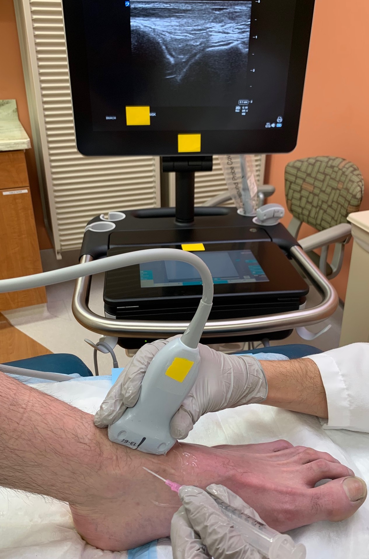
Ultrasound With Tibial Landmarks. This image illustrates the positioning and access location to the tibiotalar ankle joint with ultrasound guidance. The ultrasound monitor shows landmarks of the tibia (left) and talus (right).
Contributed by S Bartlett, DO
(Click Image to Enlarge)
Positioning and Location to Access Subtalar Ankle Joint. This image demonstrates the positioning and location for access to the subtalar ankle joint without ultrasound guidance.
Contributed by S Bartlett, DO
(Click Image to Enlarge)
Positioning and Location to Access Subtalar Ankle Joint (Lateral View). This image is a lateral view of the positioning and location for access to the subtalar ankle joint without ultrasound guidance.
Contributed by S Bartlett, DO
References
Sternbach GL, Baker FJ 2nd. The emergency joint: arthrocentesis and synovial fluid analysis. JACEP. 1976 Oct:5(10):787-92 [PubMed PMID: 1018355]
Samuelson CO Jr, Cannon GW, Ward JR. Arthrocentesis. The Journal of family practice. 1985 Feb:20(2):179-84 [PubMed PMID: 3968527]
Borzio R, Mulchandani N, Pivec R, Kapadia BH, Leven D, Harwin SF, Urban WP. Predictors of Septic Arthritis in the Adult Population. Orthopedics. 2016 Jul 1:39(4):e657-63. doi: 10.3928/01477447-20160606-05. Epub 2016 Jun 13 [PubMed PMID: 27286047]
Ungprasert P, Kaewpoowat Q, Ratapano S, Srivali N, Bischof EF Jr. Presence of crystals is not an evidence of absence of infection. The American journal of emergency medicine. 2013 Feb:31(2):455.e1-2. doi: 10.1016/j.ajem.2012.07.020. Epub 2012 Sep 1 [PubMed PMID: 22944538]
Level 3 (low-level) evidenceRoy S, Dewitz A, Paul I. Ultrasound-assisted ankle arthrocentesis. The American journal of emergency medicine. 1999 May:17(3):300-1 [PubMed PMID: 10337894]
Level 3 (low-level) evidenceBerona K, Abdi A, Menchine M, Mailhot T, Kang T, Seif D, Chilstrom M. Success of ultrasound-guided versus landmark-guided arthrocentesis of hip, ankle, and wrist in a cadaver model. The American journal of emergency medicine. 2017 Feb:35(2):240-244. doi: 10.1016/j.ajem.2016.10.056. Epub 2016 Oct 24 [PubMed PMID: 27810253]
Bruyn GA, Schmidt WA. How to perform ultrasound-guided injections. Best practice & research. Clinical rheumatology. 2009 Apr:23(2):269-79. doi: 10.1016/j.berh.2008.11.001. Epub [PubMed PMID: 19393570]
Michels F, Matricali G, Vereecke E, Dewilde M, Vanrietvelde F, Stockmans F. The intrinsic subtalar ligaments have a consistent presence, location and morphology. Foot and ankle surgery : official journal of the European Society of Foot and Ankle Surgeons. 2021 Jan:27(1):101-109. doi: 10.1016/j.fas.2020.03.002. Epub 2020 Mar 6 [PubMed PMID: 32169330]
Brockett CL, Chapman GJ. Biomechanics of the ankle. Orthopaedics and trauma. 2016 Jun:30(3):232-238 [PubMed PMID: 27594929]
Daly CH, Moake MM, Cummings ED. Point-of-Care Ultrasound-Guided Arthrocentesis of a Pediatric Septic Ankle. Pediatric emergency care. 2024 Jan 1:40(1):68-70. doi: 10.1097/PEC.0000000000003105. Epub [PubMed PMID: 38157397]
Thom C, Pozner J, Kongkatong M, Moak J. Ultrasound-Guided Talonavicular Arthrocentesis. The Journal of emergency medicine. 2021 May:60(5):633-636. doi: 10.1016/j.jemermed.2020.12.019. Epub 2021 Jan 28 [PubMed PMID: 33516576]
Ono K, Kishimoto M, Shimasaki T, Uchida H, Kurai D, Deshpande GA, Komagata Y, Kaname S. Reactive arthritis after COVID-19 infection. RMD open. 2020 Aug:6(2):. doi: 10.1136/rmdopen-2020-001350. Epub [PubMed PMID: 32763956]
Smith J, Maida E, Murthy NS, Kissin EY, Jacobson JA. Sonographically guided posterior subtalar joint injections via the sinus tarsi approach. Journal of ultrasound in medicine : official journal of the American Institute of Ultrasound in Medicine. 2015 Jan:34(1):83-93. doi: 10.7863/ultra.34.1.83. Epub [PubMed PMID: 25542943]
Smith J, Finnoff JT, Henning PT, Turner NS. Accuracy of sonographically guided posterior subtalar joint injections: comparison of 3 techniques. Journal of ultrasound in medicine : official journal of the American Institute of Ultrasound in Medicine. 2009 Nov:28(11):1549-57 [PubMed PMID: 19854970]
Gibbons RC, Zanaboni A, Genninger J, Costantino TG. Ultrasound-versus landmark-guided medium-sized joint arthrocentesis: A randomized clinical trial. Academic emergency medicine : official journal of the Society for Academic Emergency Medicine. 2022 Feb:29(2):159-163. doi: 10.1111/acem.14396. Epub 2021 Oct 23 [PubMed PMID: 34608713]
Level 1 (high-level) evidence