 Anatomy, Shoulder and Upper Limb, Anterior Humeral Circumflex Artery
Introduction
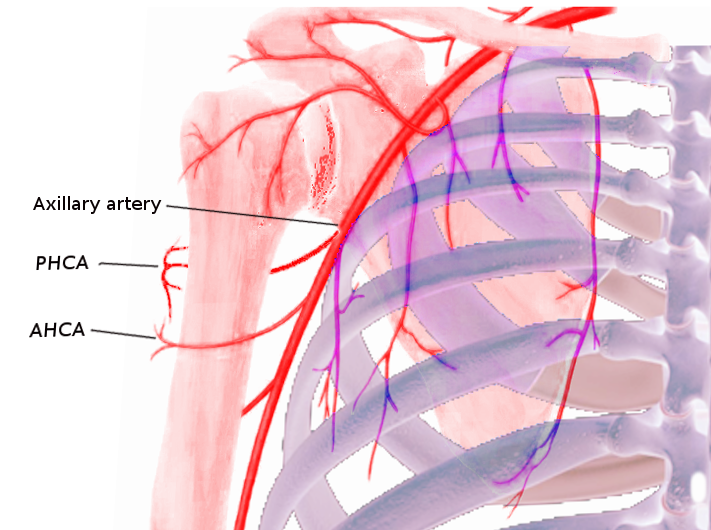
The anterior humeral circumflex artery (AHCA) branches off of the third part of the axillary artery, proximal to where the posterior humeral circumflex artery (PHCA) originates. The anterior humeral circumflex artery then travels horizontally behind the coracobrachialis muscle towards the bicipital groove (intertubercular sulcus), where its principal branch, the arcuate artery, ascends along the lateral portion of the groove and supplies the humerus at the level of the greater tuberosity. The anterior humeral circumflex artery also forms an anastomosis with the posterior humeral circumflex artery and other arterial branches, such as the profunda brachii and the acromiothoracic artery. It is crucial for the blood supply of the humeral head, the glenohumeral joint, teres major and minor, and deltoid.[1][2][3][4]
Structure and Function
Register For Free And Read The Full Article
Search engine and full access to all medical articles
10 free questions in your specialty
Free CME/CE Activities
Free daily question in your email
Save favorite articles to your dashboard
Emails offering discounts
Learn more about a Subscription to StatPearls Point-of-Care
Structure and Function
The axillary artery typically divides into three parts by the pectoralis minor muscle. The third part, which is where the anterior humeral circumflex artery originates, lies distal to the lower border of the pectoralis minor muscle. The three branches of this third part are the subscapular trunk, the anterior humeral circumflex artery, and the posterior humeral circumflex artery. The anterior humeral circumflex artery is the smallest one.[2]
It is known that the anterior humeral circumflex artery has numerous branches and an extensive anastomotic network, especially with the posterior humeral circumflex artery, in the proximal humerus. It gives off two main branches: the anterolateral ascending branch and the arcuate artery, which is the major blood supply to the greater tuberosity.[3]
The anterior humeral circumflex artery was traditionally thought to be the primary blood supply of the humerus, but recent studies suggest that the main blood supply is the posterior humeral circumflex artery. This finding may explain the relatively low rate of osteonecrosis in displaced fractures of the proximal humerus.[5][6]
Embryology
The development of the arteries of the upper limb is in close relation with upper limb development. It initiates towards the end of the fourth week with the activation of mesenchymal cells from the lateral mesoderm. This activation results in the formation of the upper limb buds, formed by a mass of mesenchymal cells covered by ectoderm. This mass of mesenchyme remains undifferentiated until later in development when it is ready to differentiate into bone, cartilage, and blood vessels.[3]
The anterior humeral circumflex artery develops from the branches of the axis artery, that comes from the lateral branch of the seventh intersegmental artery. The axis artery grows at the same rate as the upper limb, and it forms the axillary and brachial arteries from its proximal part.[7]
Blood Supply and Lymphatics
Its accompanying vein performs the venous drainage of the anterior humeral circumflex artery. The anterior circumflex humeral vein that drains into the axillary vein the majority (90%) of cases (10% of upper limbs drain into the cephalic vein).[8]
Most of the upper limb lymph nodes are located in the axilla. Based on their location, they divide into five main groups: apical, central, humeral, subscapular, and pectoral. Which sum up to about 20 to 30 nodes total. Out of these five groups, those classified as humeral are the ones most relevant to the anterior humeral circumflex artery and nearby structures. The lymphatics of the shoulder and axilla are drained by the subclavian lymphatic trunk, which continues to enter the right venous angle on the right side and is drained by the thoracic duct on the left side.[3]
Muscles
The multiple branches of the anterior humeral circumflex artery and its extensive anastomotic network (formed with posterior humeral circumflex artery) supplies oxygenated blood to the teres major, teres minor, and the deltoid muscle. As well as the long head of the biceps.[1]
Surgical Considerations
Proximal Humerus Fractures
Traditionally, surgeons have utilized a deltopectoral and a transdeltoid lateral surgical approach in open reduction and internal fixation of proximal humerus fractures. Injury to the anterior humeral circumflex artery and axillary nerve are common complications of such procedure; therefore, a couple of studies have aimed to establish anatomic landmarks to identify and protect the anterior humeral circumflex artery during surgical fixation.
The first study’s objective was to identify simple landmarks for quick access to protect both humeral circumflex arteries (anterior and posterior). Calculating the mean distances of the anterior humeral circumflex artery to the infraglenoid tubercule, the coracoid, the acromion, and the midclavicular line, which were 26.9 mm, 49.2 mm, 67.0 mm and 74.9 mm, respectively. The mean distances The second study was also aiming at identifying bony landmarks to prevent injury of the anterior humeral circumflex artery and the axillary nerve during surgical treatment, and researchers found that the anterior humeral circumflex artery was located 5.1 cm below the inferior border of the medial acromion and 2.5 cm below the prominence of the lesser tuberosity.[1][4]
Clinical Significance
Rotator Cuff Injuries
The rotator cuff is a group of four muscles: supraspinatus, subscapularis, infraspinatus, and teres minor. They arise from various points on the scapula and insert on the proximal humerus. The main function of this unit is to stabilize the glenohumeral joint, which causes them to be under great strain most of the time. Millions of Americans are affected per year by shoulder pain, most of which are caused by rotator cuff conditions. The etiology is most likely multifactorial; degeneration, overuse, and trauma. The anterior humeral circumflex artery may be associated with this condition as a cause of pain, specifically, night pain, which as a particularly troublesome symptom in patients with a rotator cuff tear, disturbing sleep, and as a result affecting the quality of life. A recent study demonstrated, using Doppler ultrasonography, that night pain was related to hemodynamics of the anterior humeral circumflex artery.[9][10][11]
Poor Posture
It is an established fact that poor posture is a significant cause of back pain, demonstrating a strong relationship with scapular kinematics and shoulder diseases. A recent cadaveric study aimed to discover how the blood flow of the anterior humeral circumflex artery changed when the scapula in internal rotation (“hunched”). It was found that the anterior humeral circumflex artery was attached to the subscapularis tendon as well as located under the subdeltoid bursa, while the posterior humeral circumflex artery was located between the deltoid muscle and the bursa. These anatomic differences determined the degree of movement during scapular motion, causing the anterior humeral circumflex artery to be more limited and the posterior humeral circumflex artery to be freely mobile. As a result, it was shown that internal rotation of the scapula decreased the blood flow of the anterior humeral circumflex artery, which might relate to several shoulder pathologies.[12]
Proximal Humerus Fractures
Proximal humerus fractures account for around 6% of fractures in the Western world. Depending on patient demographics, the fracture can occur as a result of trauma or pathologic in nature; it is the third most common osteoporotic fracture. As mentioned before, it was once thought that the anterior humeral circumflex artery was the main blood supply of the proximal humerus, but recently it has been shown that the posterior humeral circumflex artery also plays a major role. This explains the low incidence of avascular necrosis when there is a proximal humerus displaced fracture.[13]
Anterior Shoulder Dislocations
Anterior shoulder dislocations are quite common, making up to 50% of all dislocations. However, a vascular injury is a rare entity in fracture-dislocation and even more rare with isolated dislocations. The pathognomonic triad for arterial disruption includes a dislocated shoulder, diminished or absent radial pulse, and palpable axillary hematoma. These findings should prompt referral to vascular and orthopedic consult.[14]
Media
References
Chen YF, Zhu NF, Zhang CQ, Wang L, Wei HF, Lu Y. The relevance of the anatomical basis of fracture for the subsequent treatment of the anterior humeral circumflex artery and the axillary nerve. International orthopaedics. 2012 Apr:36(4):783-7. doi: 10.1007/s00264-011-1394-4. Epub 2011 Dec 24 [PubMed PMID: 22198360]
Thiel R, Munjal A, Daly DT. Anatomy, Shoulder and Upper Limb, Axillary Artery. StatPearls. 2023 Jan:(): [PubMed PMID: 29489298]
Tang A, Varacallo M. Anatomy, Head and Neck, Posterior Humeral Circumflex Artery. StatPearls. 2023 Jan:(): [PubMed PMID: 30855867]
Chen YX, Zhu Y, Wu FH, Zheng X, Wangyang YF, Yuan H, Xie XX, Li DY, Wang CJ, Shi HF. Anatomical study of simple landmarks for guiding the quick access to humeral circumflex arteries. BMC surgery. 2014 Jun 26:14():39. doi: 10.1186/1471-2482-14-39. Epub 2014 Jun 26 [PubMed PMID: 24970300]
Mostafa E, Imonugo O, Varacallo M. Anatomy, Shoulder and Upper Limb, Humerus. StatPearls. 2023 Jan:(): [PubMed PMID: 30521242]
Hettrich CM, Boraiah S, Dyke JP, Neviaser A, Helfet DL, Lorich DG. Quantitative assessment of the vascularity of the proximal part of the humerus. The Journal of bone and joint surgery. American volume. 2010 Apr:92(4):943-8. doi: 10.2106/JBJS.H.01144. Epub [PubMed PMID: 20360519]
Epperson TN, Varacallo M. Anatomy, Shoulder and Upper Limb, Brachial Artery. StatPearls. 2023 Jan:(): [PubMed PMID: 30725830]
Lee H, Bang J, Kim S, Yang H. The axillary vein and its tributaries are not in the mirror image of the axillary artery and its branches. PloS one. 2019:14(1):e0210464. doi: 10.1371/journal.pone.0210464. Epub 2019 Jan 10 [PubMed PMID: 30629680]
Terabayashi N, Watanabe T, Matsumoto K, Takigami I, Ito Y, Fukuta M, Akiyama H, Shimizu K. Increased blood flow in the anterior humeral circumflex artery correlates with night pain in patients with rotator cuff tear. Journal of orthopaedic science : official journal of the Japanese Orthopaedic Association. 2014 Sep:19(5):744-9. doi: 10.1007/s00776-014-0604-5. Epub 2014 Jul 29 [PubMed PMID: 25069807]
Level 2 (mid-level) evidenceTashjian RZ. Epidemiology, natural history, and indications for treatment of rotator cuff tears. Clinics in sports medicine. 2012 Oct:31(4):589-604. doi: 10.1016/j.csm.2012.07.001. Epub 2012 Aug 30 [PubMed PMID: 23040548]
Miniato MA, Anand P, Varacallo M. Anatomy, Shoulder and Upper Limb, Shoulder. StatPearls. 2023 Jan:(): [PubMed PMID: 30725618]
Hagiwara Y, Kanazawa K, Ando A, Nimura A, Watanabe T, Majima K, Akita K, Itoi E. Blood flow changes of the anterior humeral circumflex artery decrease with the scapula in internal rotation. Knee surgery, sports traumatology, arthroscopy : official journal of the ESSKA. 2015 May:23(5):1467-72. doi: 10.1007/s00167-013-2823-2. Epub 2014 Jan 5 [PubMed PMID: 24390057]
Schumaier A, Grawe B. Proximal Humerus Fractures: Evaluation and Management in the Elderly Patient. Geriatric orthopaedic surgery & rehabilitation. 2018:9():2151458517750516. doi: 10.1177/2151458517750516. Epub 2018 Jan 25 [PubMed PMID: 29399372]
Shah R, Koris J, Wazir A, Srinivasan SS. Anterior humeral circumflex artery avulsion with brachial plexus injury following an isolated traumatic anterior shoulder dislocation. BMJ case reports. 2016 Mar 11:2016():. doi: 10.1136/bcr-2015-213497. Epub 2016 Mar 11 [PubMed PMID: 26969353]
Level 3 (low-level) evidence