Introduction
Cutaneous angiofibroma is a benign skin tumor characterized by fibrovascular tissue and presents as a group of lesions with varied clinical appearances but consistent histological features. These benign fibrous neoplasms exhibit a proliferation of stellate and spindled cells, thin-walled blood vessels with dilated lumina in the dermis, and concentric collagen bundles. These growths typically manifest as small, firm, reddish, or flesh-colored papules, most commonly on the face (often referred to as fibrous papules or adenoma sebaceum), particularly around the nose and cheeks. However, they can also appear on other parts of the body, including the penis (as pearly penile papules), under the nails (as periungual angiofibromas or Koenen tumors), and in the mouth (as oral fibromas).
Facial angiofibromas are one of the most prominent clinical signs of tuberous sclerosis—an autosomal dominant disorder that affects the skin, kidneys, heart, brain, and lungs. In tuberous sclerosis, these angiofibromas typically emerge on the face during childhood or early adulthood (see Image. Facial Angiofibromas Observed in Tuberous Sclerosis).[1] The presence of 3 or more facial angiofibromas or 2 or more periungual angiofibromas is major diagnostic criteria for the condition. Multiple facial angiofibromas are also observed in multiple endocrine neoplasia type 1 (MEN-1) and Birt–Hogg–Dubé syndrome.[2][3][4]
Pearly penile papules are chronic, asymptomatic papules found on the coronal margin and sulcus of the penis and are more commonly observed in uncircumcised men.
Etiology
Register For Free And Read The Full Article
 Search engine and full access to all medical articles
- 10 free questions in your specialty
- Free CME/CE Activities
- Free daily question in your email
- Save favorite articles to your dashboard
- Emails offering discounts
Learn more about a Subscription to StatPearls Point-of-Care
Etiology
Tuberous sclerosis is caused by mutations in the tuberous sclerosis complex 1 (TSC 1) gene, which encodes the protein hamartin, and the tuberous sclerosis complex 2 (TSC 2) gene, which encodes the protein tuberin.[5] These proteins normally suppress the activation of the mammalian target of rapamycin (mTOR); however, when mutated, they cause unregulated proliferation of cell growth followed by the formation of multiorgan hamartomas.
MEN-1 is due to a mutation in the MEN1 gene, which encodes the protein menin. Birt-Hogg-Dubé syndrome is caused by a mutation in the FLCN gene, which encodes the protein folliculin.[6]
Epidemiology
About 75% of individuals with tuberous sclerosis eventually develop angiofibromas. Although periungual angiofibromas are less common in children, their incidence rises to 40% in adults, and they occur in 30% to 60% of patients with tuberous sclerosis. Oral fibromas are present in 30% to 70% of individuals with tuberous sclerosis, with a higher prevalence in adults than in children. Additionally, pearly penile papules are found in approximately 30% of postpubertal males.
Pathophysiology
Tuberous sclerosis is caused by mutations in the genes TSC 1, which encodes the protein hamartin, and TSC 2, which encodes the protein tuberin. These proteins normally suppress the activation of the mTOR; however, when mutated, they cause unregulated proliferation of cell growth and lead to the formation of multiorgan hamartomas. In facial angiofibromas, mTOR is activated in the proliferating fibroblast-like cells. These cells produce an epidermal growth factor called epiregulin, which stimulates epidermal cell proliferation so that they are produced at a faster rate. Additionally, angiofibromas associated with tuberous sclerosis exhibit vascular proliferation due to increased expression of angiogenic factors, such as vascular endothelial growth factor (VEGF), which further stimulates mTOR.[7][8][9]
Histopathology
All cutaneous angiofibromas comprise a dermal proliferation of fibroblasts within a collagenous stroma, accompanied by an increase in thin-walled, dilated blood vessels. Collagen fibers are arranged concentrically around hair follicles and blood vessels, while elastic fibers may be decreased, and the epidermis can be atrophic. Fibroblasts are stellate in shape and may be multinucleated. Immunohistochemistry reveals positivity for factor XIIa and negativity for S100 protein in these cells.[10]
History and Physical
Fibrous papules are solitary, dome-shaped, skin-colored to red papules typically found on the central face, particularly around the nose and malar eminences. These papules may have tiny telangiectatic vessels on their surface. In tuberous sclerosis, angiofibromas often appear symmetrically on the cheeks, nasolabial folds, nose, and chin. They may begin as erythematous macules that evolve into red or red-brown papules, which can coalesce into plaques. Rarely, angiofibromas may also be present on the scalp.[11]
Periungual angiofibromas in tuberous sclerosis typically appear from late childhood to early adulthood. They arise from the lateral or proximal nail fold, commonly affecting the toes, and can be painful, often distorting the shape of the nail. Oral fibromas most commonly occur on the gingiva but can also appear on the buccal or labial mucosa and occasionally on the tongue. Pearly penile papules are pearly, white, dome-shaped, and closely aggregated, located circumferentially on the glans penis in a multilayered manner around the corona.
Clinical findings in Birt-Hogg-Dubé syndrome include fibrofolliculomas, perifollicular fibromas (some authorities relate to angiofibromas), and trichodiscomas. These lesions typically appear as skin-colored to hypopigmented papules on the head, neck, or upper trunk.
Evaluation
The diagnosis of angiofibroma is based on history, physical examination, and skin biopsy.[10] If tuberous sclerosis, MEN-1, or Birt-Hogg-Dubé syndrome is suspected, genetic testing should be performed along with an extensive workup to identify any associated tumors according to the specific condition.
Treatment / Management
As cutaneous angiofibromas are benign lesions, they should only be removed for cosmesis or if they cause compression and pain in adjacent structures to prevent morbidity and improve outcomes. Current treatment options for angiofibromas include shave excision, cryotherapy, electrodesiccation, radiofrequency ablation, dermabrasion, and lasers such as ablative fractional laser resurfacing and pulsed dye laser. Topical podophyllotoxin is also used. Although these treatments have proven to be effective, they can result in scarring, postinflammatory hyperpigmentation, and pain.[12] (B2)
The recurrence rate of angiofibromas can be up to 80%, often requiring follow-up treatments. Topical rapamycin, an mTOR inhibitor, appears to be a safe and effective treatment for angiofibromas, although long-term studies are still needed. Combination treatments, such as fractional laser resurfacing and pulsed dye laser, can be used alongside topical medications, such as timolol or rapamycin, to effectively manage these lesions.[13][14][15](A1)
Rapamycin has recently gained popularity for treating angiofibromas. By binding to mTOR, it inhibits its activity, which reduces cell proliferation and decreases VEGF production by downregulating VEGF-stimulated endothelial cell proliferation. Several case series, case reports, and a randomized controlled trial have been published verifying the effectiveness of topical rapamycin at various concentrations and frequencies, including 0.1% used once or twice daily, 0.2% used 5 times weekly, and 0.4% used 3 times weekly. Angiofibromas typically clear while the medication is in use, with the longest reported follow-up being 3 years.
Many have used crushed rapamycin tablets mixed with Vaseline to achieve the desired concentration, though this method does not provide a standardized dose. In 2011, DeKlotz et al proposed a standardized formulation for making 0.1% topical rapamycin. Few adverse effects occur from the topical medication, primarily mild irritation and erythema. Park et al showed that topical rapamycin can effectively treat lesions smaller than 4 mm in size.[16] However, for lesions larger than 4 mm, ablative resurfacing was needed in conjunction with rapamycin. Using topical rapamycin to treat angiofibromas can be costly due to the lengthy treatment schedules required to achieve sufficient results, with expenses ranging from several hundred to several thousand dollars out of pocket.(B3)
Beta-blockers have been used for many years to treat vascular lesions. Oral propranolol has been successful in treating hemangiomas in the pediatric population, though adverse effects such as hypoglycemia limit its use in certain patients. Topical timolol 0.5% solution or gel, applied 2 to 3 times daily, has effectively treated superficial hemangiomas. The mechanism of action for beta-blockers involves blocking the conversion of renin to angiotensin II, thereby preventing the formation of VEGF, which is necessary for the conversion of endothelial stem cells to endothelial cells, thus reducing capillary development. Additionally, beta-blockers inhibit matrix metalloproteinase-9, a key enzyme in angiogenesis, and promote osteoprotegerin production, leading to apoptosis of mesenchymal and endothelial cells, further decreasing angiogenesis.[16][17](B3)
Differential Diagnosis
The differential diagnosis of cutaneous angiofibroma includes several dermatological conditions with similar clinical presentations. These include fibrous papules of the face, which are often solitary and smaller, and dermatofibromas, which are firm nodules typically found on the extremities. Basal cell carcinoma can also resemble angiofibromas but is usually more pearly and may ulcerate. Sebaceous hyperplasia presents as yellowish papules with central umbilication, often mistaken for angiofibromas. Additionally, trichoepitheliomas, benign tumors arising from hair follicles, and neurofibromas associated with neurofibromatosis should be considered.
For facial lesions, angiofibromas can be confused with acne, acrochordons, intradermal melanocytic nevi, basal cell carcinoma, and adnexal tumors. Periungual angiofibromas can resemble verruca vulgaris and subungual exostosis. Pearly penile papules can be mistaken for condyloma acuminatum and molluscum contagiosum. Accurate diagnosis often requires clinical evaluation, dermoscopy, and histopathological examination to differentiate these conditions and ensure appropriate management.
Prognosis
The prognosis of cutaneous angiofibroma is generally favorable, as these lesions are benign proliferations. While they often persist once they appear, they do not pose a significant health risk. However, when multiple, angiofibromas can cause significant disfigurement, bleeding, pruritus, and erythema, necessitating effective treatment.
Treatment for cutaneous angiofibromas is typically sought for cosmetic reasons or if the angiofibromas cause discomfort due to their size or location. Although various treatment options, such as laser therapy, cryotherapy, and topical medications like rapamycin, are effective, recurrences are common, with rates as high as 80%. Long-term management may require multiple treatments to maintain cosmetic outcomes. Overall, individuals with cutaneous angiofibromas can expect a good quality of life with appropriate care and monitoring.
Complications
Lesions in angiofibroma are prone to secondary bacterial infections and can bleed easily, causing chronic ulceration. In syndromic cases like tuberous sclerosis, angiofibromas are associated with severe systemic complications, such as renal angiomyolipomas, pulmonary lymphangioleiomyomatosis, and neurological issues, including epilepsy and cognitive impairment.
Some treatment-related complications from laser therapy or surgical excision include scarring, postinflammatory hyperpigmentation, and potential lesion recurrence. Systemic treatments, such as mTOR inhibitors, may cause immunosuppression and other adverse effects.
Deterrence and Patient Education
Patients should be educated on the importance of regular dermatologic evaluations to monitor for new lesions and manage existing ones, reducing the risk of complications such as bleeding, infection, and ulceration. Emphasis on gentle skin care and avoiding trauma to lesions can prevent secondary infections and minimize bleeding. Patients should be informed about treatment options, including laser therapy, surgical excision, and the use of topical or systemic medications such as mTOR inhibitors. They should also be made aware of potential adverse effects and the need for ongoing follow-up.
Education on the genetic nature of the condition and the potential for associated systemic complications is essential, prompting regular surveillance for renal, pulmonary, and neurological involvement in syndromic cases. Providing psychological support and resources to address the psychosocial impact of visible lesions can improve quality of life. Encouraging patients to connect with support groups and organizations can offer additional emotional support.
Enhancing Healthcare Team Outcomes
Effective management of cutaneous angiofibromas requires an interprofessional approach involving physicians, advanced practitioners, nurses, pharmacists, and other healthcare professionals. Physicians and advanced practitioners develop diagnostic skills to accurately identify cutaneous angiofibromas, differentiate them from other dermatological conditions, and master various treatment modalities such as laser therapy, cryotherapy, and topical medications.
Dermatologists and geneticists address the complex needs of patients, especially those with underlying genetic conditions such as tuberous sclerosis. Nurses provide wound care and patient education to support posttreatment recovery and adherence to management plans. Pharmacists offer expertise in dermatological pharmacotherapy, particularly in the use of topical treatments such as rapamycin, to ensure safe and effective medication use.
Treatment plans must be tailored to individual patient needs, considering both medical and cosmetic outcomes to enhance quality of life. The team collaboratively monitors treatment efficacy and potential recurrences, adjusting the management plan as necessary. By fostering a collaborative, informed, and patient-centered approach, healthcare professionals can significantly enhance the management of cutaneous angiofibromas, leading to improved clinical outcomes, increased safety, and better team performance.
Media
(Click Image to Enlarge)
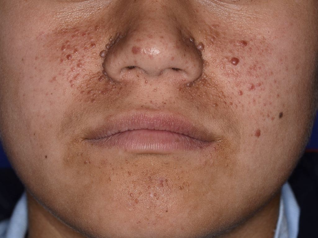
Facial Angiofibromas Observed in Tuberous Sclerosis. Facial angiofibromas are considered one of the most apparent clinical presentations of tuberous sclerosis, an autosomal dominant hamartomatous disorder that affects the skin, kidneys, heart, brain, and lungs.
References
Bakić M, Ratković M, Gledović B, Vujović B, Radunović D, Babić V, Prelević V. Cutaneous Manifestations of Tuberous Sclerosis. Acta dermatovenerologica Croatica : ADC. 2018 Apr:26(1):73-74 [PubMed PMID: 29782307]
Pierotti L, Pardi E, Dinoi E, Piaggi P, Borsari S, Della Valentina S, Sardella C, Michelucci A, Caligo MA, Bogazzi F, Marcocci C, Cetani F. Cutaneous lesions and other non-endocrine manifestations of Multiple Endocrine Neoplasia type 1 syndrome. Frontiers in endocrinology. 2023:14():1191040. doi: 10.3389/fendo.2023.1191040. Epub 2023 Jul 7 [PubMed PMID: 37484956]
Kundu GK, Ahmed S, Akhter S, Islam MT, Dwa T, Sabbir AM. Clinical and Neuro-radiological Presentation of Tuberous Sclerosis Complex in Tertiary Care Centre. Mymensingh medical journal : MMJ. 2018 Jan:27(1):144-148 [PubMed PMID: 29459606]
Wang S, Liu Y, Wei J, Zhang J, Wang Z, Xu Z. Tuberous Sclerosis Complex in 29 Children: Clinical and Genetic Analysis and Facial Angiofibroma Responses to Topical Sirolimus. Pediatric dermatology. 2017 Sep:34(5):572-577. doi: 10.1111/pde.13204. Epub 2017 Aug 2 [PubMed PMID: 28771801]
Amorim BDB, Azulay D, Ramos-E-Silva M. Tuberous Sclerosis. Skinmed. 2021:19(3):179-185 [PubMed PMID: 34303387]
Adam MP, Feldman J, Mirzaa GM, Pagon RA, Wallace SE, Bean LJH, Gripp KW, Amemiya A, Sattler EC, Steinlein OK. Birt-Hogg-Dubé Syndrome. GeneReviews(®). 1993:(): [PubMed PMID: 20301695]
Garcia NG, de Carli ML, Oliveira DT, Soares CT, Ribeiro Júnior NV, Sperandio FF, Hanemann JA. Tuberous Sclerosis with Severe Cutaneous Manifestation and Multiples Facial Angiofibromas. Head and neck pathology. 2016 Dec:10(4):542-546 [PubMed PMID: 27140177]
DiCicco B, Johnson W, Allred J, Soldano AC, Ramsdell WM. Koenen's tumor and facial angiofibromas in a case of Birt-Hogg-Dubé syndrome: A cutaneous contribution to growing evidence of a relationship with tuberous sclerosis complex. JAAD case reports. 2016 May:2(3):196-8. doi: 10.1016/j.jdcr.2016.03.014. Epub 2016 May 25 [PubMed PMID: 27274535]
Level 3 (low-level) evidenceBouguéon G, Lagarce F, Martin L, Pailhoriès H, Bastiat G, Vrignaud S. Formulation and characterization of a 0.1% rapamycin cream for the treatment of Tuberous Sclerosis Complex-related angiofibromas. International journal of pharmaceutics. 2016 Jul 25:509(1-2):279-284. doi: 10.1016/j.ijpharm.2016.05.064. Epub 2016 Jun 1 [PubMed PMID: 27260135]
Nasimi M, Kamyab K, Moradi A, Dasdar S, Kianfar N. Clinical and histopathological evaluation of cutaneous angiofibromas. Journal of cutaneous pathology. 2021 Oct:48(10):1262-1265. doi: 10.1111/cup.14060. Epub 2021 Jun 4 [PubMed PMID: 33977570]
Freitas LF, Segundo LBM, Costa DMC, Duarte ML, Tibana LAT. Atypical cutaneous presentation of tuberous sclerosis complex: Giant angiofibroma on the scalp. Arquivos de neuro-psiquiatria. 2022 Apr:80(4):450-451. doi: 10.1590/0004-282X-ANP-2021-0315. Epub [PubMed PMID: 34932647]
Boggarapu S, Roberds SL, Nakagawa J, Beresford E. Characterization and management of facial angiofibroma related to tuberous sclerosis complex in the United States: retrospective analysis of the natural history database. Orphanet journal of rare diseases. 2022 Sep 14:17(1):355. doi: 10.1186/s13023-022-02496-2. Epub 2022 Sep 14 [PubMed PMID: 36104799]
Level 2 (mid-level) evidenceSalomon FF, Barreto MM, Zanetti G, Rodrigues RS, Gasparetto EL, Marchiori E. CNS and cutaneous involvement in tuberous sclerosis complex. Arquivos de neuro-psiquiatria. 2015 Sep:73(9):813. doi: 10.1590/0004-282X20150101. Epub [PubMed PMID: 26352504]
Wataya-Kaneda M, Tanaka M, Yang L, Yang F, Tsuruta D, Nakamura A, Matsumoto S, Hamasaki T, Tanemura A, Katayama I. Clinical and Histologic Analysis of the Efficacy of Topical Rapamycin Therapy Against Hypomelanotic Macules in Tuberous Sclerosis Complex. JAMA dermatology. 2015 Jul:151(7):722-30. doi: 10.1001/jamadermatol.2014.4298. Epub [PubMed PMID: 25692384]
Level 1 (high-level) evidenceMa G, Wu P, Lin X, Chen H, Li W, Hu X, Jin Y. Nd:YAG laser for "fractional" treatment of angiofibromas. International journal of dermatology. 2014 May:53(5):638-42. doi: 10.1111/ijd.12384. Epub 2014 Mar 6 [PubMed PMID: 24602020]
Park J, Yun SK, Cho YS, Song KH, Kim HU. Treatment of angiofibromas in tuberous sclerosis complex: the effect of topical rapamycin and concomitant laser therapy. Dermatology (Basel, Switzerland). 2014:228(1):37-41. doi: 10.1159/000357033. Epub 2013 Dec 21 [PubMed PMID: 24401865]
Level 3 (low-level) evidencePatel AM, Chou EL, Findeiss L, Kelly KM. The horizon for treating cutaneous vascular lesions. Seminars in cutaneous medicine and surgery. 2012 Jun:31(2):98-104. doi: 10.1016/j.sder.2012.02.001. Epub [PubMed PMID: 22640429]