Introduction
Cephalometric analysis evaluates lateral skull radiographs obtained with a cephalostat to determine skeletal patterns and assess treatment complexity. This technique is specifically indicated for cases involving planned anteroposterior movements but is not necessary for all orthodontic treatments. The use of cephalometric analysis is particularly justified when significant modifications to the incisor position are anticipated.
The technique of cephalometric analysis has a rich history, originating in the late 1800s when radiographs were first used to study the head and neck. In the 1930s, Holly Broadbent, a professor of orthodontics at the University of Michigan, advanced the field by analyzing the relationship between the teeth and the skull. This pioneering work involved measuring various angles and distances on radiographic images, thereby establishing the foundations of cephalometric analysis.[1] Over the following decades, researchers expanded on this foundational work, developing additional methods such as the Wits analysis. Cephalometric analysis is an integral part of modern orthodontics practice, aiding in the diagnosis and treatment of various dental and skeletal anomalies.[2]
Anatomy and Physiology
Register For Free And Read The Full Article
 Search engine and full access to all medical articles
- 10 free questions in your specialty
- Free CME/CE Activities
- Free daily question in your email
- Save favorite articles to your dashboard
- Emails offering discounts
Learn more about a Subscription to StatPearls Point-of-Care
Anatomy and Physiology
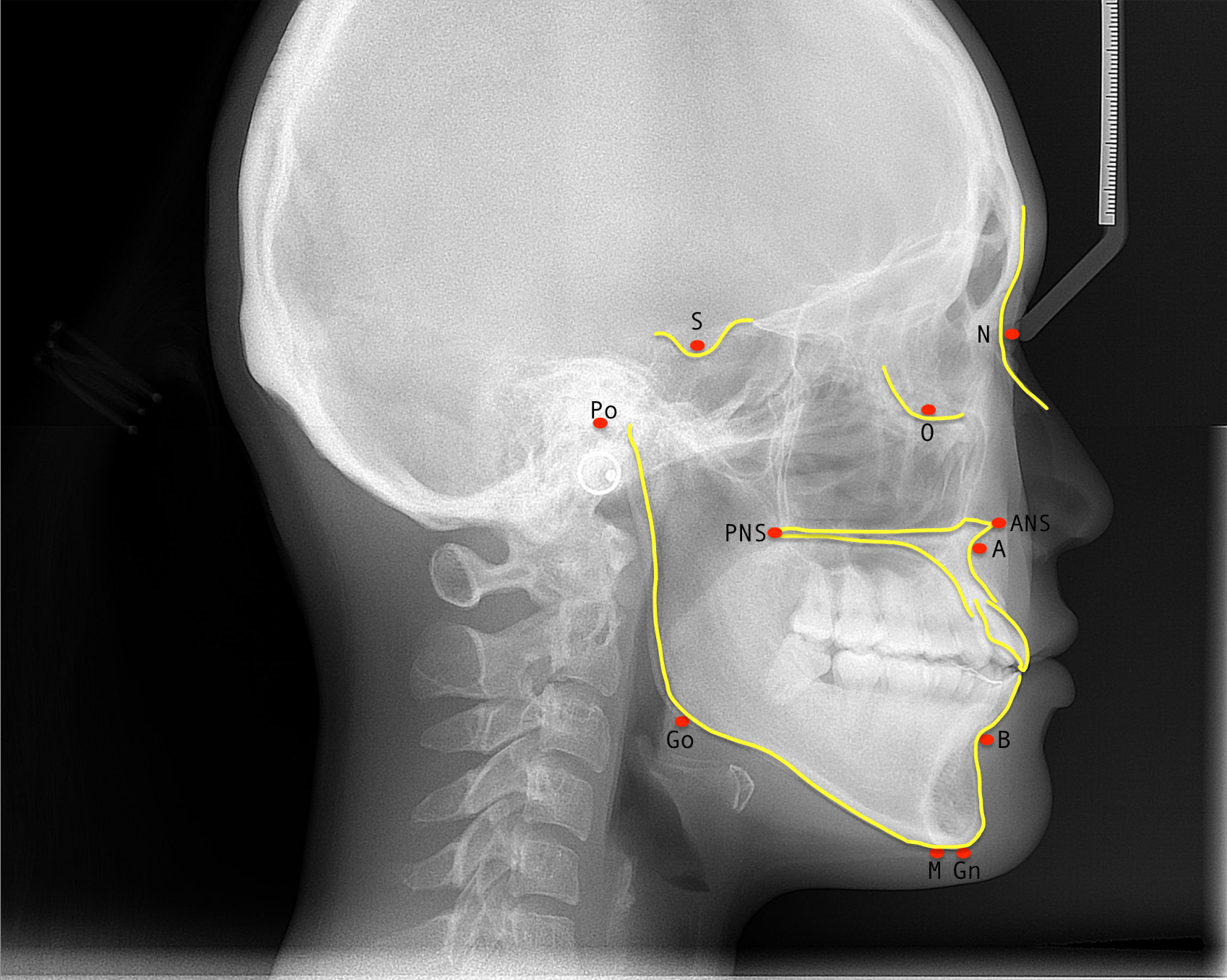
Cephalometric analysis assesses the anteroposterior and vertical relationships of the mandible and maxilla in relation to the cranial base and each other, as well as the upper and lower teeth in relation to the mandibular and maxillary bones (see Image. Common Cephalometric Points). The analysis involves comparing patient-specific proportions and angle measurements with population averages, which can be obtained through manual tracing or digital methods. The results of the cephalometric analysis are subject to projection errors and measurement variability and are dependent on the operator. Therefore, the cephalometric values for an individual patient must be interpreted in the clinical context, as deviations from average values may be compensated by other features in the face or skull. The tables below describe commonly used cephalometric points (see Table 1) and planes (see Table 2).[2][3]
Table 1. Cephalometric Points
| Cephalometric Points | Anatomical Description |
| Sella (S) | The midpoint of the sella turcica. |
| Nasion (N) | The most anterior point of the frontonasal suture or the deepest point of the intersection of the frontal and nasal bones. |
| Orbitale (Or) | The most anteroinferior point on the infraorbital margin, averaged from the right and left shadows. |
| Porion (Po) | The most superior and external point of the bony external auditory meatus or the level of the superior border of the mandibular condyle. |
|
Anterior nasal spine (ANS) |
The tip of the ANS. |
| Posterior nasal spine (PNS) | The tip of the PNS. |
| A-point (A) | The most posterior point of the concavity of the maxilla, located between the ANS and the maxillary alveolar process. |
| B-point (B) | The most posterior point of the concavity of the mandible, located between the pogonion and the crest of the mandibular alveolar process. |
| Menton (Me) | The most inferior point on the mandibular symphysis in the midline. |
| Pogonion (Pog) | The most anterior point on the contour of the bony chin. |
| Gnathion (Gn) | The lowest and most forward point on the chin outline. |
| Condylion (Cd) | The lowest and most superoposterior point on the curvature of the average of the right and left outlines of the condylar heads. |
Table 2. Cephalometric Planes
| Cephalometric Planes | Anatomical Description |
| Sella-Nasion (SN) | A horizontal plane passing through the sella turcica and nasion; used as a reference for measuring the anteroposterior relationship between the jaws and facial structures. |
| Frankfort | A horizontal plane formed by a line that joins the porion and orbitale; used as a reference for assessing the vertical relationship between the jaws and facial structures. |
| Mandibular | A line through the inferior border of the mandible, joining the gonion and menton; used as a reference for evaluating the vertical position of the mandible. |
| Maxillary | A line joining the anterior and posterior nasal spines; used as a reference for assessing the angulation and position of the maxilla. |
| Occlusal | A horizontal plane touching the incisal edges of the maxillary and mandibular incisors and the tips of the occlusal surfaces of the posterior teeth. |
| Camper | A horizontal plane passing through the inferior margin of the nasal ala and the superior margin of the tragus; used as a reference for evaluating the inclination of the upper lip and the position of the incisors. |
Indications
Cephalometric analysis helps diagnose dental and skeletal malocclusions, plan corrective treatments, and evaluate treatment outcomes and growth changes.
Technique or Treatment
Standardized Lateral Cephalometric Radiographs
Lateral skull radiographs provide a 2-dimensional representation of the head and neck and measure the sagittal and vertical dimensions of the skull. Sagittal measurements assess the position and inclination of the maxilla and mandible, while vertical measurements evaluate the height of the facial structures and the relationship between the jaw.[4] In contrast, posteroanterior radiographs, taken from the front of the head, measure transverse and vertical dimensions, offering insights into the width of the face and the relationship between the jaws in the transverse plane.[5] However, in clinical practice, cephalometric analysis primarily relies on lateral radiographs, as posteroanterior projections are more challenging to interpret.
A standardized technique is essential for obtaining radiographs to ensure accurate comparisons over time and between patients. Image quality depends heavily on the proper positioning of the patient. The patient should be positioned with the Frankfort plane horizontal, ear rests placed in the external auditory meatuses, the nasion aligned on the bridge of the nose, and teeth in centric occlusion.[6] The radiographic source is fixed at a distance of 5 feet (150–180 cm) from the patient's mid-sagittal plane, with a film-to-mid-sagittal plane distance of 30 cm.[6] A calibrated steel ruler is included in each image, ensuring precise measurements are documented.
Cephalometric Tracing
Lateral cephalometric radiographs are traditionally traced manually. First, a tracing acetate is placed over the film. Anatomical landmarks are identified and marked on the acetate using a sharp 4H pencil. These points are then connected to form lines and angles, and the resulting measurements are recorded and interpreted.
Digital tracing using specialized software streamlines the cephalometric analysis process. The software automatically identifies anatomical landmarks on radiographs and calculates measurements. This also provides standards for comparison based on ethnicity, sex, and age while enabling predictions for soft tissue alterations, growth, and surgical outcomes.[7] Both manual and digital tracing techniques are appropriate for cephalometric analysis.
Clinical Significance
Interpretation of Cephalometric Analysis Results
Average angular measurements and proportions are established for the general population, but these values serve as general guidelines. Standard measures may vary based on age, sex, and ethnicity.[8] A comprehensive orthodontic assessment must incorporate the patient’s unique skeletal and dental characteristics alongside cephalometric findings.
Anteroposterior Evaluation
SNA angle: The SNA angle assesses the anteroposterior position of the maxilla relative to the anterior cranial base.[9] The SNA angle is formed by connecting the sella, nasion, and A-point. The average SNA angle is 81 ± 3 degrees. For example, a patient with an SNA angle of 82 degrees demonstrates a well-positioned maxilla relative to the cranial base.[10]
An increased SNA angle indicates that the maxilla is positioned protrusively relative to the cranial base compared to the average.[3] Conversely, a decreased SNA angle suggests that the maxilla is retruded relative to the cranial base compared to the norm.[3]
SNB angle: The SNB angle evaluates the anteroposterior position of the mandible to the anterior cranial base.[9] The SNB angle is formed by connecting the sella, nasion, and B-point. The average SNB angle is 78 ± 3 degrees.[10] An increased SNB angle indicates that the mandible is positioned anteriorly relative to the cranial base compared to the average.[11] Conversely, a decreased SNB angle suggests that the mandible is retruded relative to the cranial base compared to the norm.[11]
ANB angle: The ANB angle evaluates the anteroposterior relationship between the maxilla and mandible.[12] The ANB angle is calculated as the difference between the SNA (sella-nasion to A-point) and SNB (sella-nasion to B-point) angles, using the formula: ANB = SNA - SNB.[10]
The average ANB angle for a class I skeletal pattern is 2 degrees. An ANB angle greater than 4 degrees suggests a class II skeletal pattern, while an angle less than 2 degrees indicates a class III skeletal pattern.[13] However, the ANB angle can vary based on the position of the nasion and the prominence of the lower face. When the ANB angle is abnormally increased or decreased, alternative methods such as the Wits analysis should be considered.
Wits Analysis
The Wits analysis provides an alternative approach to evaluating the anteroposterior skeletal pattern without relying on the cranial base. This method involves drawing perpendicular lines from points A and B to the occlusal plane, defined as the line joining the cusps of the posterior teeth.[14] The intersections of the perpendiculars from points A and B with the occlusal plane define points AO and BO, respectively.[14] The distance between AO and BO is then measured. For a class I skeletal pattern, BO is typically 1 mm (±1.9 mm) anterior to AO in males, while in females, BO and AO are generally equal (±1.77 mm).[14]
Vertical Evaluation
The maxillary-mandibular plane angle (MMPA) assesses the vertical relationship between the maxilla and mandible. The MMPA is formed by projecting lines from the mandibular and maxillary planes until they meet posteriorly. The average MMPA value is 27 ± 4 degrees. A normal MMPA indicates a well-proportioned lower face and a normal overbite. An increased MMPA value suggests a long lower face and an open bite, while a decreased MMPA value indicates a shorter lower face and a closed bite.[15]
Incisor Position: Angular Evaluation
The angular measurement of the maxilla is determined by measuring the angle from the incisor to nasion-A, while the angular measurement of the mandible is calculated from the incisor to nasion-B. These values indicate the inclination of teeth—proclined teeth are angled forward, retroclined teeth are tilted backward, and normally inclined teeth are in a neutral position.[8] The normal incisor to nasion-A angle is 22 degrees, and the normal incisor to nasion-B angle is 25 degrees. An increased incisor to nasion-A or nasion-B angle indicates proclination of the incisor, while a decreased angle suggests retroclination.[3]
The position of mandibular incisors is further evaluated by the angle formed between the long axis of the tooth and the mandibular plane, which extends from gonion to gnathion.[10] The normal mandibular incisor to mandibular plane angle (Go-Gn) is 87 degrees. An increased mandibular incisor to mandibular plane angle indicates that the incisors are proclined, whereas a decreased value indicates the incisors are retroclined.[8]
Incisor Position: Linear Evaluation
The linear measurement of the maxilla is taken from the maxillary incisor to nasion-A, while the linear measurement of the mandible is taken from the mandibular incisor to nasion-B. These measurements describe the relationship between the tooth and its supporting basal bone, indicating whether the tooth is in a normal, procumbent (ahead of the supporting bone), or recumbent (behind the supporting bone) position.[10]
The average value for both incisor to nasion-A and incisor to nasion-B is 4 mm. An increased incisor to nasion-A or -B value indicates that the incisor is procumbent, whereas a decreased value suggests that the incisor is recumbent.[16]
Enhancing Healthcare Team Outcomes
Cephalometric analysis is crucial in diagnosing and treating malocclusions, requiring an interprofessional healthcare team of dental health professionals, including general dentists, orthodontists, and oral surgeons. This technique provides valuable insights into the extent of skeletal and dental misalignments, as well as potential causative factors. If a malocclusion is too severe to be treated by an orthodontist alone, a referral to an oral surgeon may be necessary. The oral surgeon can collaborate with the orthodontist to correct the misaligned jaw through orthognathic surgery, thereby highlighting the importance of an interprofessional approach to diagnosing and managing complex orthodontic malocclusions. Meticulous planning and collaboration with other healthcare professionals involved in orthodontic treatment are essential for ensuring successful patient outcomes.
Media
(Click Image to Enlarge)
References
Jha MS. Cephalometric Evaluation Based on Steiner's Analysis on Adults of Bihar. Journal of pharmacy & bioallied sciences. 2021 Nov:13(Suppl 2):S1360-S1364. doi: 10.4103/jpbs.jpbs_172_21. Epub 2021 Nov 10 [PubMed PMID: 35017989]
Hans MG, Palomo JM, Valiathan M. History of imaging in orthodontics from Broadbent to cone-beam computed tomography. American journal of orthodontics and dentofacial orthopedics : official publication of the American Association of Orthodontists, its constituent societies, and the American Board of Orthodontics. 2015 Dec:148(6):914-21. doi: 10.1016/j.ajodo.2015.09.007. Epub [PubMed PMID: 26672697]
Bergman RT. Cephalometric soft tissue facial analysis. American journal of orthodontics and dentofacial orthopedics : official publication of the American Association of Orthodontists, its constituent societies, and the American Board of Orthodontics. 1999 Oct:116(4):373-89 [PubMed PMID: 10511665]
Level 3 (low-level) evidenceAldrees AM. Lateral cephalometric norms for Saudi adults: A meta-analysis. The Saudi dental journal. 2011 Jan:23(1):3-7. doi: 10.1016/j.sdentj.2010.09.002. Epub 2010 Oct 1 [PubMed PMID: 24151411]
Level 1 (high-level) evidenceDinesh A, Mutalik S, Feldman J, Tadinada A. Value-addition of lateral cephalometric radiographs in orthodontic diagnosis and treatment planning. The Angle orthodontist. 2020 Sep 1:90(5):665-671. doi: 10.2319/062319-425.1. Epub [PubMed PMID: 33378477]
Gupta S, Tandon P, Singh GP, Shastri D. Comparative assessment of cephalometric with its analogous photographic variables. National journal of maxillofacial surgery. 2022 Jan-Apr:13(1):99-107. doi: 10.4103/njms.NJMS_267_20. Epub 2022 Apr 20 [PubMed PMID: 35911811]
Level 2 (mid-level) evidenceTsorovas G, Karsten AL. A comparison of hand-tracing and cephalometric analysis computer programs with and without advanced features--accuracy and time demands. European journal of orthodontics. 2010 Dec:32(6):721-8. doi: 10.1093/ejo/cjq009. Epub 2010 Jun 16 [PubMed PMID: 20554891]
Scheideman GB, Bell WH, Legan HL, Finn RA, Reisch JS. Cephalometric analysis of dentofacial normals. American journal of orthodontics. 1980 Oct:78(4):404-20 [PubMed PMID: 6933849]
Brevi B, Di Blasio A, Di Blasio C, Piazza F, D'Ascanio L, Sesenna E. Which cephalometric analysis for maxillo-mandibular surgery in patients with obstructive sleep apnoea syndrome? Acta otorhinolaryngologica Italica : organo ufficiale della Societa italiana di otorinolaringologia e chirurgia cervico-facciale. 2015 Oct:35(5):332-7. doi: 10.14639/0392-100X-415. Epub [PubMed PMID: 26824915]
Tenti FV. Cephalometric analysis as a tool for treatment planning and evaluation. European journal of orthodontics. 1981:3(4):241-5 [PubMed PMID: 6945994]
Schwendicke F, Chaurasia A, Arsiwala L, Lee JH, Elhennawy K, Jost-Brinkmann PG, Demarco F, Krois J. Deep learning for cephalometric landmark detection: systematic review and meta-analysis. Clinical oral investigations. 2021 Jul:25(7):4299-4309. doi: 10.1007/s00784-021-03990-w. Epub 2021 May 27 [PubMed PMID: 34046742]
Level 1 (high-level) evidenceDi Blasio A, Di Blasio C, Pedrazzi G, Cassi D, Magnifico M, Manfredi E, Gandolfini M. Combined photographic and ultrasonographic measurement of the ANB angle: a pilot study. Oral radiology. 2017:33(3):212-218. doi: 10.1007/s11282-017-0275-y. Epub 2017 Mar 21 [PubMed PMID: 28890606]
Level 3 (low-level) evidencePlaza SP, Reimpell A, Silva J, Montoya D. Relationship between skeletal Class II and Class III malocclusions with vertical skeletal pattern. Dental press journal of orthodontics. 2019 Sep 5:24(4):63-72. doi: 10.1590/2177-6709.24.4.063-072.oar. Epub 2019 Sep 5 [PubMed PMID: 31508708]
Jacobson A. The "Wits" appraisal of jaw disharmony. American journal of orthodontics. 1975 Feb:67(2):125-38 [PubMed PMID: 1054214]
Kotuła J, Kuc AE, Lis J, Kawala B, Sarul M. New Sagittal and Vertical Cephalometric Analysis Methods: A Systematic Review. Diagnostics (Basel, Switzerland). 2022 Jul 15:12(7):. doi: 10.3390/diagnostics12071723. Epub 2022 Jul 15 [PubMed PMID: 35885628]
Level 1 (high-level) evidenceLentini-Oliveira DA, Carvalho FR, Rodrigues CG, Ye Q, Prado LB, Prado GF, Hu R. Orthodontic and orthopaedic treatment for anterior open bite in children. The Cochrane database of systematic reviews. 2014 Sep 24:2014(9):CD005515. doi: 10.1002/14651858.CD005515.pub3. Epub 2014 Sep 24 [PubMed PMID: 25247473]
Level 1 (high-level) evidence