Continuing Education Activity
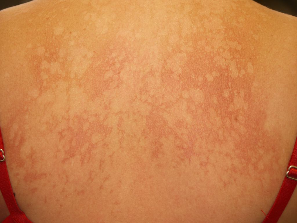
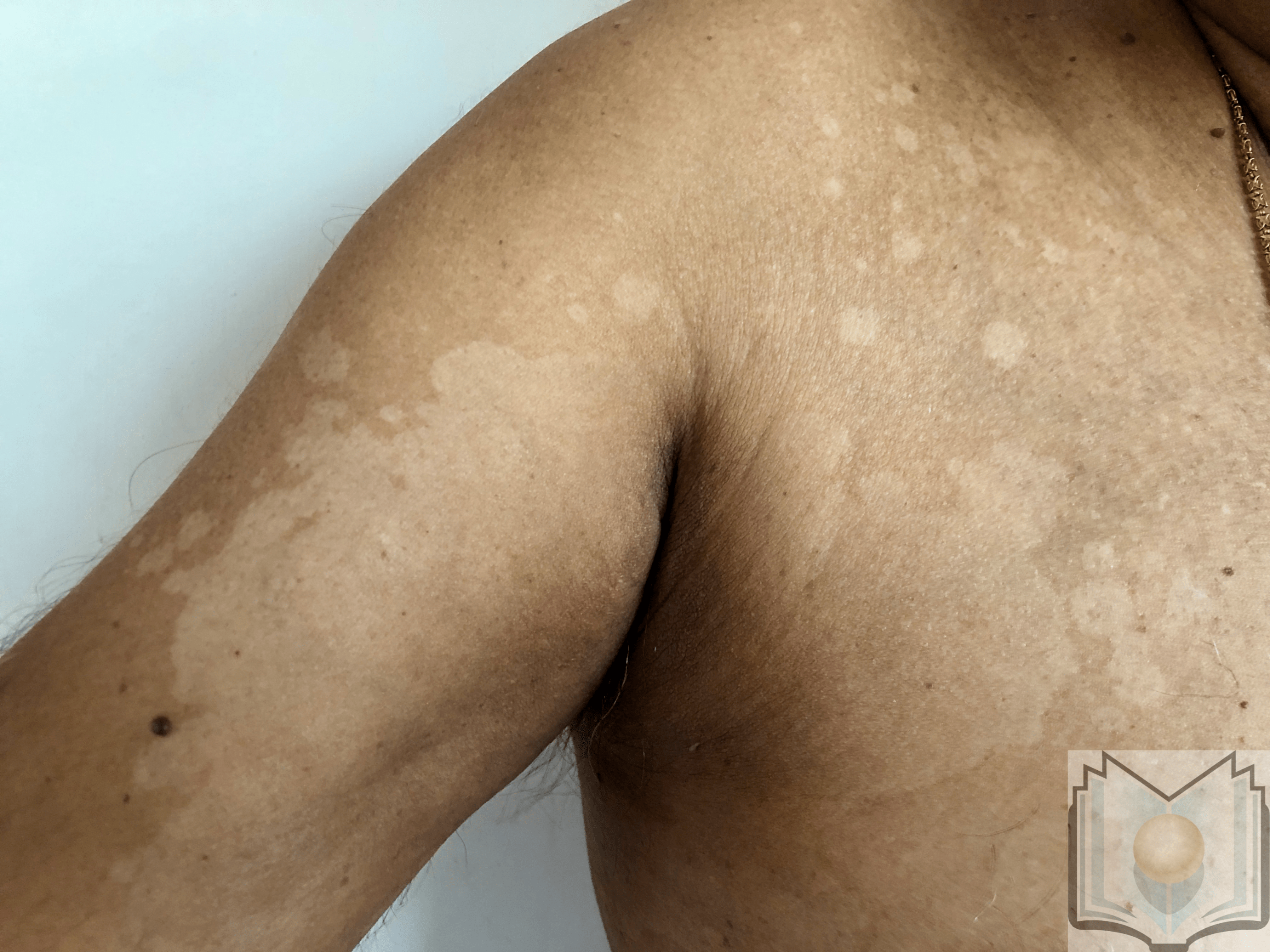
Pityriasis versicolor, also known as tinea versicolor, is a common, benign, superficial fungal infection of the skin. Clinical features of pityriasis versicolor include either hyperpigmented or hypopigmented finely scaled macules. The most frequently affected sites are the trunk, neck, and proximal extremities. This activity reviews the evaluation and management of pityriasis versicolor and highlights the role of interprofessional team members in collaborating to provide well-coordinated care and enhance patient outcomes.
Objectives:
- Describe the epidemiology of pityriasis versicolor.
- Explain the evaluation for pityriasis versicolor.
- Outline treatment considerations for pityriasis versicolor.
- Summarize the importance of enhancing care coordination among the interprofessional team to ensure proper evaluation and management of pityriasis versicolor.
Introduction
Pityriasis versicolor, also known as tinea versicolor, is a frequent, benign, superficial fungal infection of the skin. It belongs to Malassezia-related diseases. Clinical features of pityriasis versicolor include either hyperpigmented or hypopigmented finely scaly macules. The most frequently affected sites are the trunk, neck, and proximal extremities.[1][2][3] The diagnosis of pityriasis versicolor is often made on clinical grounds alone. The ultraviolet black light and the microscopic examination of scales soaked in potassium hydroxide may be helpful in doubtful cases. Pityriasis versicolor responds well to induction therapy. However, long-term maintenance treatment often is required because of high recurrence rate.
Etiology
Pityriasis versicolor is caused by Malassezia, a dimorphic lipophilic fungus, also known as Pityrosporum. It is a component of normal skin flora. To date, 14 species of Malassezia have been identified. [3]The main species isolated in pityriasis versicolor are Malassezia furfur, Malassezia globosa, Malassezia sympodialis.
Epidemiology
Pityriasis versicolor has been reported worldwide, but it is more common in warm and humid conditions. The prevalence is as high as 50% in tropical countries and as low as 1.1% in cold climates such as Sweden. Pityriasis versicolor occurs more frequently in adolescents and young adults probably due to the increase of sebum production by the sebaceous glands which allow for a more lipid-rich environment in which Malassezia can grow. Pityriasis versicolor affects men and women equally and no specific ethnic predominance has been noted.[4][5]
Pathophysiology
Malassezia is commensal of healthy skin, and it is most common in oily areas such as the face, scalp, and back. However, Malassezia can cause pityriasis versicolor when it converts to its pathogenic filamentous form. Factors that lead to this pathogenic conversion include a genetic predisposition, environmental conditions such as heat and humidity, immunodeficiency, pregnancy, oily skin, and application of oily lotions and creams.
Histopathology
A skin biopsy is not required to confirm a diagnosis, but if it is performed, histological findings include hyperkeratosis, acanthosis, and a mild superficial, perivascular infiltrate in the dermis. Fungal elements are localized almost exclusively within the stratum corneum and can often be visualized even in sections stained with hematoxylin-eosin. Both spores and hyphae of Malassezia are present and are often likened to spaghetti and meatballs. Periodic acid-Schiff stain may improve recognition of the fungus.
History and Physical
Patients with pityriasis versicolor present with multiple, well-demarcated, oval, finely scaling patches or plaques. Skin lesions may be hypopigmented, hyperpigmented, or erythematous and occasionally become confluent and widespread. The fine scale may not be readily apparent on the lesions, but it is easily provoked when the affected skin is stretched or scraped. The distribution of affected skin reflects the lipophilic nature of the fungus since the seborrheic areas (trunk, neck, and/or arms) are predominantly involved. The face also may be affected, particularly in children. Pityriasis versicolor skin lesions are usually asymptomatic or slightly pruritic. However, severe pruritus can be present in very warm and humid conditions.
Evaluation
Diagnosis of pityriasis versicolor is usually easily made on the basis of its characteristic clinical presentation (hyperpigmented or hypopigmented, finely scaling patches or plaques).[6][7][8]
The ultraviolet black light (Wood lamp) may help to demonstrate the coppery-orange fluorescence of pityriasis versicolor.
The diagnosis is confirmed by microscopic examination of scales soaked in potassium hydroxide examination, which demonstrates the typical grape-like clusters of yeast cells and long hyphae. Since the standard potassium-hydroxide mount lacks a color contrast, methylene blue stain, ink blue stain, or Swartz-Medrik stain may be added to visualize better.
Malassezia species are known to be difficult to grow in the laboratory as they require fastidious culture conditions.
Treatment / Management
Patients should be informed that the causative agent of pityriasis versicolor is a commensal fungal inhabitant of the normal skin flora, and therefore the disease is not considered to be contagious. In addition, pityriasis versicolor does not lead to either permanent scarring or pigmentary disorders. However, in many cases, disease recurrence may occur despite effective treatment.[9][10][11]
Pityriasis versicolor may be treated effectively with topical and/or systemic agents.
Topical medications are considered the first-line therapy for pityriasis versicolor. Topical treatments are divided into nonspecific antifungal agents (sulfur plus salicylic acid, selenium sulfide 2.5%, and zinc-pyrithione) that primarily remove dead tissue and prevent further invasion, and specific antifungal drugs, that have fungicidal or fungistatic effects. Antifungal agents include imidazole (clotrimazole 1%, ketoconazole 2%, econazole, isoconazole, miconazole), ciclopirox olamine 1% , and allylamine (terbinafine 1%). Galenic forms such as sprays or foaming solutions in shampoo are preferable to creams because creams are oilier and more difficult to apply, especially in widespread areas. Ketoconazole is the most common topical treatment used to treat pityriasis versicolor. It can be applied as a cream (twice daily for 15 days) or in a foaming solution (single dose).
Oral medications are viewed as a second-line of treatment for pityriasis versicolor in the event of widespread, severe, recalcitrant or recurrent cases. Systemic therapies include itraconazole (200 mg daily for seven days) and fluconazole (150 to 300 mg weekly dose for 2 to 4 weeks) that are preferred to oral ketoconazole which is no longer approved due to its potential hepatotoxic sides effects. Oral terbinafine is not effective in the treatment of pityriasis versicolor.
In cases of recurrent pityriasis versicolor, maintenance therapy may be necessary. Topical prophylactic treatment can be used. However, systemic antifungal agents are preferred since they are less time-consuming and ensure better compliance. Many studies have been conducted to identify the best prophylactic regimen.
Differential Diagnosis
Pityriasis versicolor may be confused with various conditions:
- Pityriasis rosea
- Tinea corporis
- Vitiligo
- Pityriasis alba
- Confluent reticulated papillomatosis of Gougerot and Carteaud
- Postinflammatory hypo- and hyperpigmentation
- Seborrheic dermatitis
- Guttate psoriasis
- Tinea corporis
- Nummular eczema
- Secondary syphilis
- Mycosis fungoides
Prognosis
Pityriasis versicolor is benign and noncontagious since the causative fungal pathogen is commensal of normal skin. Oral and topical antifungal agents are effective; however, disease recurrence is common and may have an impact on a patient's quality of life. Therefore, preventive measures should be taken. Also, patients must be reminded that pigmentary changes may take weeks to months to clear, even if the fungus is eradicated.
Pearls and Other Issues
Pityriasis versicolor is a benign but commonly recurrent superficial fungal infection of the skin. Therefore, patients need effective follow-up care to implement a relapse prevention strategy.
Enhancing Healthcare Team Outcomes
Tinea versicolor is a relatively common skin disorder that may be encountered by the nurse practitioner, internist, dermtologist and primary care physician. The diagnosis is usually made on clinical features but does require clinical acumen. The rash is benign and may spontaneously disappear.
Patients should be informed that the causative agent of pityriasis versicolor is a commensal fungal inhabitant of the normal skin flora, and therefore the disease is not considered to be contagious. In addition, pityriasis versicolor does not lead to either permanent scarring or pigmentary disorders. However, in many cases, disease recurrence may occur despite effective treatment. An interprofessional team of a nurse and clinician should provide patient education which will decrease their anxiety and provide the best patient outcome.
The outcomes in most patients are excellent.[12][13] (Level V)