Continuing Education Activity
Metacarpal fractures account for 40% of all hand fractures. A fracture of the neck of the fifth metacarpal, or boxer's fracture, named for the classic mechanism of injury in which direct trauma is applied to a clenched fist, is the most common, representing 10% of all hand fractures. Treatment for a boxer's fracture varies based on whether the fracture is open or closed, the degree of angulation, rotation, and other concomitant injuries. Immobilization with an ulnar gutter splint may be the definitive treatment for closed, non-displaced fractures without angulation or rotation, while open fractures, significantly angulated or malrotated fractures or those involving injury to neurovascular structures require referral to a hand surgeon. This activity reviews the etiology, presentation, evaluation, and management of boxer's fracture, and reviews the role of the interprofessional team in evaluating, diagnosing, and managing the condition.
Objectives:
- Describe the mechanism of injury that results in a boxer's fracture of the hand.
- Review the necessary elements for an examination to assess for boxer's fracture, including any necessary diagnostic imaging studies.
- Summarize the treatment options available for fractures of the neck of the fifth metacarpal, including both conservative and surgical care.
- Explain the importance of improving care coordination among the interprofessional team to enhance the delivery of care for patients with fifth metacarpal neck fractures.
Introduction
Metacarpal fractures account for 40% of all hand fractures. [1] A Boxer’s fracture is a fracture of the fifth metacarpal neck, named for the classic mechanism of injury in which direct trauma is applied to a clenched fist. This represents 10% of all hand fractures. [1]Treatment for a Boxer’s fracture varies based on whether the fracture is open or closed, characteristics of the fracture including the degree of angulation, shortening, and rotation, and other concomitant injuries. Immobilization with an ulnar gutter splint may be the definitive treatment for closed, non-displaced fractures without angulation or rotation, while open fractures, significantly angulated or malrotated fractures or those involving injury to neurovascular structures may require operative fixation.
Etiology
The most common mechanism of injury for Boxer’s fracture is punching, e.g., the axial pressure applied to the metacarpal bone when the fist is in a clenched position. Direct trauma to the dorsum of the hand may also cause a fracture of the fifth metacarpal neck. Unlike many other hand and wrist fractures, a Boxer’s fracture typically does not occur with a fall onto an outstretched hand.
Epidemiology
The incidence of metacarpal neck fractures presenting for hospital care in the United States is 13.6 per 100,000 person-years.[1] Metacarpal fractures account for 40% of all hand fractures, [[1] while fractures of the fifth metacarpal neck account for 10% of all hand fractures.[1] The incidence in males is five times higher than in females.[2] Males aged ten to 19 have the highest incidence, followed by males aged 20 to 29[2].They commonly occur at home and at sporting/athletic events [2], [3].
Pathophysiology
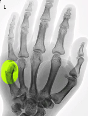
The fifth metacarpal bone is one of the five metacarpal bones of the hand. The fifth metacarpal is associated with the fifth digit. The metacarpal bone consists of a head (distally located), neck, body, and base (proximally located). Axial load via direct trauma to a clenched fist transfers energy to the metacarpal bone, causing fractures most commonly at the neck, and typically resulting in apex dorsal angulation due in part to the forces exerted by the pull of the interosseous muscles. The interosseous muscles, responsible for adduction and abduction of the fingers, originate from the metacarpal shafts and insert onto proximal phalanges. The collateral ligaments also join the metacarpal bones to the proximal phalanges and must be taken into consideration during splinting to minimize the risk of loss of motion due to shortening of the ligaments. The ligaments are taut in flexion, and more slack in extension, therefore the MCP joints should be splinted in flexion to prevent shortening (intrinsic plus positioning)[4]. The arteries and nerves supplying the fingers are adjacent to the metacarpal bones and can be injured in severely displaced Boxer’s fractures, requiring surgical intervention.
History and Physical
Patients with Boxer’s fractures present with complaints of dorsal hand pain, swelling, and possible deformity in the setting of one of the mechanisms typically associated with this injury involving direct trauma to the hand.
Complete physical exam of a potential Boxer’s fracture should include an examination of the entire hand, comparison to the contralateral hand, with special attention to the following:
- Skin: Closely inspect the skin for any breaks, especially near the metacarpal head, typically the point of impact. When a Boxer’s fracture is sustained by a blow to the face, the recipient’s tooth may cause a laceration or abrasion known as a “fight bite." This may require operative irrigation and debridement.
- Neurovascular exam: As with all suspected fractures, a neurovascular exam should test for sensation, motor function, and blood flow distal to the injury.
- Angulation: Boxer’s fractures are typically associated with apex dorsal angulation, thereby resulting in depression of the MCP joint and loss of the normal knuckle contour. With significantly angulated fractures, “pseudo-clawing” may be observed due to damage to the extensor apparatus; pseudo-clawing is a hyperextension of the MCP joint and flexion at the PIP joint. The degree of angulation is determined using plain films.
- Rotational alignment: Any degree of malrotation warrants referral to a hand surgeon and therefore assessment of rotational alignment is a crucial component of the physical exam. Alignment can be assessed by examining the hand with the MCP and PCP joints in flexion, and DIP joints extended. If lines are drawn along the digits and extended distally, normally aligned digits will show the convergence of these lines. If the line extended from the fifth finger does not converge towards the others, suspect malrotation.
- Malrotation can also be detected by examining the hand with the MCPs flexed, and PCPs and DIPs extended. The fingernails should be in line along a single plane.
Evaluation
Plain radiographs are the standard of care to diagnose Boxer’s fractures and determine a degree of angulation. Anteroposterior, lateral, and oblique views should be obtained. The lateral view should be used to measure the degree of angulation of the shaft of the metacarpal as compared to the mid-point of the fracture fragment.[5] Normal angulation of the metacarpal head to the neck is 15 degrees, so the angulation of the fracture should be measured as that more than the baseline of 15 degrees.
Recent literature suggests that bedside ultrasound may also be used to make an initial diagnosis of a Boxer’s fracture[6].
CT is generally not used for the diagnosis of metacarpal fractures; however occult fractures may be detected via CT in patients for whom there is a high degree of clinical suspicion for fracture and negative plain radiographs[7].
Treatment / Management
The appropriate treatment for a Boxer’s fracture on initial presentation varies based on whether the fracture is open or closed, the degree of angulation, rotation, and other concomitant injuries. Due to the risk of infection from "fight bite," even very small wounds should be thoroughly irrigated, and there should be a low threshold for antibiotic treatment.
Immobilization Alone
For a Boxer’s fracture that is closed, not angulated, and not malrotated or otherwise displaced, splinting is used for initial immobilization. A Boxer’s fracture should be immobilized with an ulnar gutter splint. Alternatively, a pre-made Galveston splint or a custom orthosis may be used.
The hand should be positioned in the intrinsic plus position for splinting: mild wrist extension, 70 to 90 degrees of flexion at MCP joint, and slight flexion at the DIP and PIP joints. Flexion of these joints is important to prevent shortening of the collateral ligaments and subsequent loss of range of motion and functional impairment.
Closed Reduction
Closed reduction is required for a Boxer’s fracture with significant angulation greater than 30 degrees.
Analgesia options for the procedure include a hematoma block or an ulnar nerve block. Younger children or very anxious patients may require procedural sedation, but this procedure typically is tolerated well without sedation.
Closed reduction of a Boxer’s fracture is accomplished by using the “90-90 method.” The MCP, DIP, and PIP joints should all be flexed to 90 degrees. The clinician should then apply volar pressure over the dorsal aspect of the fracture site while applying pressure axially to the flexed PIP joint. This axial pressure to the PIP applies dorsal force to the distal fracture fragment. The clinician should be able to feel the reduction when it has been achieved. The injury should be immobilized with an ulnar gutter splint, and post-reduction films should be taken to assess for adequate reduction [8]. The fifth metacarpal neck can tolerate angulation of up to 50-60 degrees and management may be continued non-operatively if remains within the acceptable tolerances.
Surgical Referral
Surgical referral is indicated for fractures that are open, severely comminuted, associated with neurovascular injury, and for fractures with any malrotation[9]. Surgical referral is also appropriate for fractures with significant angulation if the initial provider is unsuccessful in achieving adequate reduction and alignment outside acceptable parameters. Surgical options include open reduction internal fixation, or closed reduction percutaneous pinning.
Follow-Up
Boxer’s fractures should be sent for repeat radiographs within one week to assess alignment. Radiographs should be obtained every two weeks following, until clinical and radiographic healing are present, typically between four to six weeks. Even with the adequate reduction, some cosmetic deformity may persist, with loss of the normal knuckle contour. After a short period of immobilization, the passive and active range of motion exercises should be performed to alleviate stiffness of the MCP and PIP joints. Literature supports early mobilization of these injuries rather than prolonged immobilization[10], [11]. If any loss of function persists after several weeks of these exercises, referral to occupational therapy is warranted.
Differential Diagnosis
Differential diagnosis for 5th metacarpal neck fractures include: fractures involving the metacarpal head, shaft, and base.
Prognosis
Literature has shown that closed management of fifth metacarpal neck fractures with less than 60-70 degrees of angulation have high functional capabilities as indicated by quickDASH scores at four months [12].
Complications
Complications of fifth metacarpal neck fractures include digit malrotation, nonunion, and loss of esthetic appears of the fifth knuckle [12]. These should be communicated with the patient when discussing operative and non-operative treatments.
Deterrence and Patient Education
If patients have minimal pain and no impairment of function, conservative treatment may be sufficient, even for angulated fractures. However, if there is any evidence of severe angulation, comminuted fractures, neurovascular injury, functional impairment, or unmanageable pain, patients should be referred to a hand surgeon. Thorough education with patients regarding management options should be discussed.
Enhancing Healthcare Team Outcomes
The delayed presentation is not uncommon with Boxer’s fracture, possibly due to some hesitation to present for care given the classic mechanism of the injury. Clinicians and nurses must be aware of and work together to identify patients with his injury. When patients present 2 to 3 weeks after sustaining this injury, as with initial presentation, assess function, angulation, and pain; these fractures typically heal without any functional detriment [Level V].