Autosomal Recessive Polycystic Kidney Disease

Earn CME/CE in your profession:


Continuing Education Activity

There are two inherited varieties of polycystic kidney disease (PKD): autosomal dominant polycystic kidney disease (ADPKD) and autosomal recessive polycystic kidney disease (ARPKD). These two forms have distinct clinical and genetic features. ADPKD is a multisystem progressive cystic disorder characterized by bilateral renal cysts arising anywhere along the nephron and leading to progressively worsening fibrosis and architectural distortion of kidneys and ultimately renal failure. ARPKD involves microcystic cysts that that arise exclusively in the collecting tubule and can lead to hepatic fibrosis and consequently, manifestations of portal hypertension. This activity reviews the evaluation, treatment, and complications of PKD and the importance of an interprofessional team approach to its management.

Objectives:

  • Identify the etiologies of polycystic kidney disease.

  • Describe the presentations of polycystic kidney disease.

  • Review the treatment options for polycystic kidney disease.

  • Summarize the importance of optimizing coordination amongst the interprofessional team to enhance the delivery of care to patients with polycystic kidney disease.

Introduction

There are two varieties of polycystic kidney disease based on inheritance: autosomal dominant (ADPKD) and autosomal recessive (ARPKD) types. These two forms have distinct clinical and genetic features.

ADPKD is a multisystem progressive cystic disorder. It is characterized by bilateral renal cysts, which progressively lead to fibrosis and architectural distortion of kidneys, and progressive renal failure.[[1] Other organs, such as the liver, pancreas, and spleen, can also be involved.  These patients also have a higher risk of developing aneurysms along the circle of Willis than the general population. In adult patients, it is the most frequent genetic cause of renal failure. It accounts for 5% of patients on dialysis in the United States.[[2] Nearly 50% of patients with ADPKD progresses to end-stage renal disease by the age of 65 years.[3]

ARPKD primarily involves the kidney and liver. Historically referred to as infantile polycystic kidney disease, with the current knowledge about the genetic basis of the disease and clinical manifestation, it can present as infantile, juvenile, or even in the adult population. Hence old nomenclature of adult and infantile polycystic kidney disease is not used anymore.[4] It is characterized by renal collecting duct ectasia, hepatic biliary duct ectasia/malformation, and fibrosis involving both the liver and kidney. 

Cystic dilatation of renal tubules characterizes both ADPKD and ARPKD. ADPKD shows cysts of varying sizes which may show coarse calcifications and renal calculi. Cysts in ARPKD are mostly microcystic. Cysts in ADPKD can arise from anywhere along the nephron, most commonly from the collecting tube. However, cysts in ARPKD arise exclusively in the collecting tubule.

Etiology

ADPKD is the most common hereditary renal cystic disease.[5] The condition may arise from mutation of either of two genes, PKD 1 and PKD 2. PKD 1 located in the short arm of chromosome 16p and encodes protein polycystic-1 which accounts for a majority of cases (85%). PKD 2 is located on the long arm of chromosome 4q and encodes protein polycystic-2, which accounts for the remaining 15% of cases.[6]

There is considerable variability in the phenotypic expression of ARPKD. All forms of ARPKD are caused by a mutation in the PKHD1 gene on chromosome 6p12.[7] There is evidence of extensive alternative splicing of this gene. It is known that a critical amount of full-length protein secreted by this gene is responsible for the normal function of tubular epithelium. These mutations can present in the prenatal period, infancy, childhood, or early adulthood period.

Epidemiology

ADPKD occurs in 1 in 400-1000 live births, without any sex or racial predilection.[5] Each child of the affected individual has a 50% chance of inheriting the mutations with complete penetrance. About 5% of cases occur due to spontaneous mutations.[8]

ARPKD is one of the most common causes of heritable, infantile cystic renal disease. Despite that, it accounts for only 1 in 20,000 to 50,000 live births.[9] There is no gender or racial predilection.

Pathophysiology

Variable expression of PKD 1 and PKD 2 genes causes ADPKD. PKD 1 gene-disease has more severe manifestations than PKD 2 genes. Polycystin 1 and 2 proteins are components of cell membranes of primary cilia of renal tubular epithelial cells.[10] They play a very critical role in cellular physiology by regulating intracellular calcium transport.[11] Deranged production of these proteins results in ciliary dysmotility, which leads to overproduction of epidermal growth factors, which causes proliferation of tubular epithelial cells with increased fluid secretion and cysts formation.[12]

Cyclic adenosine monophosphate, a cellular second messenger, also induces the proliferation of tubular epithelial cells. Mutations also cause mislocalization of sodium-potassium-activated adenosine triphosphate at the apical membrane of tubular epithelial cells, resulting in secretion of sodium and a gradient formation accounting for fluid secretion and cyst formation. According to Knudson's two-hit hypothesis, all the renal tubular epithelial cells of the patient carry a germline mutation in PKD1 and PKD2. When somatic mutations occur in the normal allele, a second hit takes place. This leads to the monoclonal proliferation of that cell causes further pathogenesis of the disease.[13] Hepatic cysts arise by excessive proliferation and dilatation of biliary ductules, caused by a similar mechanism.

Intrarenal cysts distort the normal renal architecture and alter function, causing continued activation of the renin-angiotensin-aldosterone system which leads to hypertension.[14] 

Pathogenesis of ARPKD is characterized by circumferential proliferation of epithelial cells, which predominantly affects collecting ducts in renal tubules.[15] They cause variable lengthening and ectasia of renal tubules. The abnormal proliferation of renal tubule epithelium causes them to lose their normal physiological function. They begin to secrete fluid in the ducts. The fluid is rich in epithelial growth factors, which leads to further proliferation of epithelial cells.[16] Animal ARPKD model studies on epithelial growth receptor blockers and epithelial enzymes have shown promising results, which may pave the way for therapeutic interventions at the cellular level.[16]

Histopathology

ADPKD: Gross, histologic specimen reveals bilaterally enlarged kidneys with multiple cysts of varying sizes. On microscopy, renal tubular ectasia and cysts lined by columnar to cuboidal to flattened epithelial cells and thickened basement membrane are the usual findings.[5]

ARPKD: Variable degrees of collecting duct epithelial proliferation, biliary duct proliferation and ectasia, periportal fibrosis are present in almost all patients.[17]

History and Physical

ADPKD: Patient may present clinically usually around the third decade of age with hypertension and with or without pain or hematuria of renal origin or other cyst's related-complications as infection or rupture.

Renal stones are a known complication of ADPKD, which contributes to significant morbidity. Medical renal disease as renal tubular acidosis is also common in this population. About 45% of patients may progress to end-stage renal disease, which leads to their dependence on hemodialysis or renal transplant. Imaging studies play a vital role in the diagnosis and screening of families at risk.

ARPKD: Early identification of these patients has increased tremendously in the last few decades with routine antenatal ultrasound which usually shows large and echogenic kidneys. The majority present in the neonatal period with hypertension and renal dysfunction. Hepatic fibrosis may lead to manifestations of portal hypertension. There is a variable phenotypic presentation of the disease depending on the severity of renal and hepatic involvement, which usually have an inverse relationship.

Perinatal Type is the most common. It presents with oligohydramnios and pulmonary hypoplasia and has a poor prognosis.

Neonatal and Infantile Type: Minimal to moderate hepatic/periportal fibrosis.

Juvenile Type: Gross hepatic fibrosis and features of portal hypertension like hepatosplenomegaly and portosystemic varices with less severe renal disease.

Evaluation

ADPKD: 

Imaging of patients with ADPKD can be tedious because of the enlarged, distorted kidneys and variably sized cysts and their mass effects on adjacent structures.

Ultrasound is cheap, easy, fast, and lacking in ionizing radiation, and is an excellent choice for regular follow-up of these cases. Simple cysts appear anechoic on ultrasound, while complex cysts may show anechoic material and thick septa or calcifications, which require careful assessment to rule out hemorrhage, infection, or renal malignancy. Ultrasound is also used to assess cysts in other organs like the liver.

CT is a very sensitive tool in imaging renal cysts. Any complex cysts showing thick enhancing septa and a solid nodule are viewed with caution and classified according to the Bosniac classification.

MRI is as informative as CT with the added benefit of lack of ionizing radiation. Simple cysts appear hyperintense on T2 and hypointense on T1. Hemorrhage in a cyst appears hyperintense on T1; calcification shows blooming on GRE or susceptibility-weighted images. A complex cyst may show thick enhancing septa or enhancing solid nodules which raise suspicion for malignancy.

Recently,  image-based renal and cyst volumetry measurement has been implemented in several institutions and utilized as an indicator for the assessment of treatment response or disease progression.[18] MR-based imaging is beneficial in such volumetric assessment. Patients with ADPKD are known to have a mean renal volume of more than 1,000ml (average normal value is 150 ml) with an age-related increase in renal and cystic volumes. Assessment of glomerular filtration rate (GFR) by nuclear imaging is usually done to gain further information on renal function.[18][19] Data from the Consortium for Radiologic Imaging of Polycystic Kidney Disease (CRISP) suggest that renal structure (volume) and function (GFR) share an inverse relationship and are directly proportional to the development of hypertension as well as urinary albumin excretion in individuals with normal renal function.[20]

ARPKD:

In the perinatal and neonatal period, ultrasound typically shows bilateral smooth enlarged kidneys with loss of corticomedullary differentiation. CT shows smooth enlarged kidneys with a striated pattern of contrast excretion.[21] The striated pattern on CT signifies a collection of contrast in dilated renal tubules. 

Pulmonary hypoplasia resulting from oligohydramnios caused by the perinatal manifestation of ARPKD can be a cause of significant mortality and morbidity in such patients.[22] The liver usually appears normal in these neonates. However, very few may develop dilated intrahepatic biliary ducts.[23]

Those children who survive the neonatal period and present later with portal hypertension in the infantile or juvenile period usually have less involvement of kidneys. Their renal ultrasound is usually normal or may show minimal cysts.[24] Other findings which may be associated with ARPKD include; biliary duct ectasia (Caroli disease) and congenital hepatic fibrosis.[25][26] A subset group of ARPKD patients may show hepatosplenomegaly.[27]

Genetic testing of an affected sibling with ARPKD is another tool in the evaluation of these families and patients at risk.[25][26]  The absence of renal findings in the patient's biological parents increases the suspicion of ARPKD and prompts further genetic testing.[26]

Treatment / Management

The treatment of patients with ADPKD includes managing high blood pressure with medications, a low-salt diet, dietary protein restriction, and statins which may reduce disease progression.[28]

Selected patients may receive treatment with a vasopressin receptor antagonist, tolvaptan. Few randomized clinical trials have shown convincing results of tolvaptan in checking the progression of kidney disease in ADPKD, but it remains the only FDA-approved therapy for ADPKD.[29][30]

Targeted therapies for hereditary renal cystic diseases are undergoing extensive clinical studies. Drugs targeting mTOR signaling pathways, like rapamycin, by checking cellular proliferation are under Phase II/III clinical trials. A variety of drugs like methylprednisone, urine alkalinization, lovastatin, epidermal growth factor tyrosine kinase receptor inhibitor, and cyclin-dependent kinase inhibitor are undergoing animal studies to assess utility in this group of patients.[31][32][33]

For example, combined somatostatin and tolvaptan block the effect of cyclic adenosine monophosphate and inhibit fluid secretion and cell proliferation. Triptolide, which affects calcium signaling, also exhibits antiproliferative effects. A number of other agents may prove helpful in halting the progression of autosomal dominant polycystic kidney disease.

Patients with ADPKD who ultimately progress to end-stage renal disease require renal replacement therapy, which includes dialysis and kidney transplantation. 

ARPKD: Management depends on the severity of the clinical manifestations and the organs involved; this involves monitoring respiratory function, renal function tests, liver function tests, infant growth evaluation and blood pressure monitoring, and symptomatic treatment. Dual organ transplant (liver and kidney), depending on the severity of portal hypertension and end-stage renal disease, has shown promising results in a significant number of cases.[34]

Finally, genetic counseling is of the paramount value for both patients and families.

Differential Diagnosis

Differentials for ADPKD:

  • Acquired renal cystic disease
  • Autosomal dominant tubulointerstitial kidney disease (formerly called medullary cystic kidney disease)
  • Developmental renal cystic disease- medullary sponge kidney, multicystic dysplastic kidney disease
  • Localized renal cystic disease
  • Tuberous sclerosis complex
  • Von Hippel Lindau disease

Differentials for ARPKD:

  • ADPKD
  • Beckwith-Wiedemann Syndrome
  • Laurence-Moon-Beidl-syndrome
  • Meckel-Gruber syndrome
  • Renal dysplasia associated with trisomy 13 (Patau syndrome)

Prognosis

ADPKD: In a majority of patients, the renal function remains intact until the fourth decade of life. Once renal function is affected, glomerular filtration rate (GFR) starts to decline, at an average rate of 4.4 to 5.9 mL/min per year.[1] Other than end-stage renal disease, most patients with ADPKD die from cardiac causes.[35]

ARPKD: Prognosis is poor. Prognosis depends on the severity of renal disease. Neonates born with severe renal disease may not survive due to pulmonary hypoplasia and insufficiency.[36]. Those with less severe disease may survive the neonatal period and develop progressive renal failure and end-stage renal disease.

The liver disease in ARPKD is relatively mild in neonatal and early infancy. The severity of disease is known to progress with age. These children develop features of portal hypertension because of chronic liver fibrosis. However as portal hypertension and variceal bleeding are not life-threatening if properly managed, many of these patients survive up to middle age.

Complications

Complications related to renal structure and functions as discussed above are possible.

Deterrence and Patient Education

Patients and their families must receive education regarding their condition, disease progression, complications, and management options. Social support to patient and families along the course of treatment and management are the recommendation.

Enhancing Healthcare Team Outcomes

An interprofessional approach and management are essential in the treatment of patients with polycystic kidney disease. Patients may need regular follow up with a neurologist, gastroenterologist, dietitian, nephrologist, radiologist, and pediatrician. Lifelong monitoring for complications is necessary.


(Click Image to Enlarge)
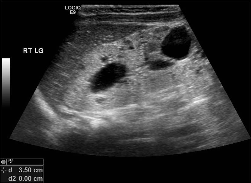
ARPKD in a 6-month -old girl. Enalrged and ecogenic kidney with cysts.
Contributed by Surabhi Subramanian .

(Click Image to Enlarge)
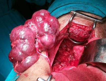
Polycystic kidney disease
Contributed by Sunil Munakomi, MD
Details

Editor:

Tahani Ahmad

Updated:

11/4/2023 12:09:53 PM

References


[1]

Torres VE, Harris PC. Autosomal dominant polycystic kidney disease: the last 3 years. Kidney international. 2009 Jul:76(2):149-68. doi: 10.1038/ki.2009.128. Epub 2009 May 20     [PubMed PMID: 19455193]


[2]

Lentine KL, Xiao H, Machnicki G, Gheorghian A, Schnitzler MA. Renal function and healthcare costs in patients with polycystic kidney disease. Clinical journal of the American Society of Nephrology : CJASN. 2010 Aug:5(8):1471-9. doi: 10.2215/CJN.00780110. Epub 2010 Jun 10     [PubMed PMID: 20538839]


[3]

Akoh JA. Current management of autosomal dominant polycystic kidney disease. World journal of nephrology. 2015 Sep 6:4(4):468-79. doi: 10.5527/wjn.v4.i4.468. Epub     [PubMed PMID: 26380198]


[4]

Guay-Woodford LM, Galliani CA, Musulman-Mroczek E, Spear GS, Guillot AP, Bernstein J. Diffuse renal cystic disease in children: morphologic and genetic correlations. Pediatric nephrology (Berlin, Germany). 1998 Apr:12(3):173-82     [PubMed PMID: 9630032]


[5]

Truong LD, Choi YJ, Shen SS, Ayala G, Amato R, Krishnan B. Renal cystic neoplasms and renal neoplasms associated with cystic renal diseases: pathogenetic and molecular links. Advances in anatomic pathology. 2003 May:10(3):135-59     [PubMed PMID: 12717117]

Level 3 (low-level) evidence

[6]

Bisceglia M, Galliani CA, Senger C, Stallone C, Sessa A. Renal cystic diseases: a review. Advances in anatomic pathology. 2006 Jan:13(1):26-56     [PubMed PMID: 16462154]

Level 3 (low-level) evidence

[7]

Bergmann C, Senderek J, Küpper F, Schneider F, Dornia C, Windelen E, Eggermann T, Rudnik-Schöneborn S, Kirfel J, Furu L, Onuchic LF, Rossetti S, Harris PC, Somlo S, Guay-Woodford L, Germino GG, Moser M, Büttner R, Zerres K. PKHD1 mutations in autosomal recessive polycystic kidney disease (ARPKD). Human mutation. 2004 May:23(5):453-63     [PubMed PMID: 15108277]


[8]

Grantham JJ. Clinical practice. Autosomal dominant polycystic kidney disease. The New England journal of medicine. 2008 Oct 2:359(14):1477-85. doi: 10.1056/NEJMcp0804458. Epub     [PubMed PMID: 18832246]


[9]

Traubici J, Daneman A. High-resolution renal sonography in children with autosomal recessive polycystic kidney disease. AJR. American journal of roentgenology. 2005 May:184(5):1630-3     [PubMed PMID: 15855129]


[10]

Zhang Q, Taulman PD, Yoder BK. Cystic kidney diseases: all roads lead to the cilium. Physiology (Bethesda, Md.). 2004 Aug:19():225-30     [PubMed PMID: 15304637]


[11]

Fliegauf M, Benzing T, Omran H. When cilia go bad: cilia defects and ciliopathies. Nature reviews. Molecular cell biology. 2007 Nov:8(11):880-93     [PubMed PMID: 17955020]


[12]

Wilson PD, Goilav B. Cystic disease of the kidney. Annual review of pathology. 2007:2():341-68     [PubMed PMID: 18039103]


[13]

Berger AH, Knudson AG, Pandolfi PP. A continuum model for tumour suppression. Nature. 2011 Aug 10:476(7359):163-9. doi: 10.1038/nature10275. Epub 2011 Aug 10     [PubMed PMID: 21833082]


[14]

Li X, Wüthrich RP, Kistler AD, Rodriguez D, Kapoor S, Mei C. Blood Pressure Control for Polycystic Kidney Disease. Polycystic Kidney Disease. 2015 Nov:():     [PubMed PMID: 27512778]


[15]

Martinez JR, Grantham JJ. Polycystic kidney disease: etiology, pathogenesis, and treatment. Disease-a-month : DM. 1995 Nov:41(11):693-765     [PubMed PMID: 7587886]


[16]

Richards WG, Sweeney WE, Yoder BK, Wilkinson JE, Woychik RP, Avner ED. Epidermal growth factor receptor activity mediates renal cyst formation in polycystic kidney disease. The Journal of clinical investigation. 1998 Mar 1:101(5):935-9     [PubMed PMID: 9486961]


[17]

Kamath BM, Piccoli DA. Heritable disorders of the bile ducts. Gastroenterology clinics of North America. 2003 Sep:32(3):857-75, vi     [PubMed PMID: 14562578]


[18]

Chapman AB. Approaches to testing new treatments in autosomal dominant polycystic kidney disease: insights from the CRISP and HALT-PKD studies. Clinical journal of the American Society of Nephrology : CJASN. 2008 Jul:3(4):1197-204. doi: 10.2215/CJN.00060108. Epub 2008 Jun 25     [PubMed PMID: 18579674]


[19]

Grantham JJ, Torres VE, Chapman AB, Guay-Woodford LM, Bae KT, King BF Jr, Wetzel LH, Baumgarten DA, Kenney PJ, Harris PC, Klahr S, Bennett WM, Hirschman GN, Meyers CM, Zhang X, Zhu F, Miller JP, CRISP Investigators. Volume progression in polycystic kidney disease. The New England journal of medicine. 2006 May 18:354(20):2122-30     [PubMed PMID: 16707749]


[20]

Chapman AB, Guay-Woodford LM, Grantham JJ, Torres VE, Bae KT, Baumgarten DA, Kenney PJ, King BF Jr, Glockner JF, Wetzel LH, Brummer ME, O'Neill WC, Robbin ML, Bennett WM, Klahr S, Hirschman GH, Kimmel PL, Thompson PA, Miller JP, Consortium for Radiologic Imaging Studies of Polycystic Kidney Disease cohort. Renal structure in early autosomal-dominant polycystic kidney disease (ADPKD): The Consortium for Radiologic Imaging Studies of Polycystic Kidney Disease (CRISP) cohort. Kidney international. 2003 Sep:64(3):1035-45     [PubMed PMID: 12911554]


[21]

Gleason DC, McAlister WH, Kissane J. Cystic disease of the kidneys in children. The American journal of roentgenology, radium therapy, and nuclear medicine. 1967 May:100(1):135-46     [PubMed PMID: 4960727]


[22]

Sweeney WE Jr, Avner ED. Diagnosis and management of childhood polycystic kidney disease. Pediatric nephrology (Berlin, Germany). 2011 May:26(5):675-92. doi: 10.1007/s00467-010-1656-1. Epub 2010 Oct 29     [PubMed PMID: 21046169]


[23]

Chilton SJ, Cremin BJ. The spectrum of polycystic disease in children. Pediatric radiology. 1981:11(1):9-15     [PubMed PMID: 7254933]


[24]

Levine E, Hartman DS, Meilstrup JW, Van Slyke MA, Edgar KA, Barth JC. Current concepts and controversies in imaging of renal cystic diseases. The Urologic clinics of North America. 1997 Aug:24(3):523-43     [PubMed PMID: 9275977]


[25]

Hartung EA, Guay-Woodford LM. Autosomal recessive polycystic kidney disease: a hepatorenal fibrocystic disorder with pleiotropic effects. Pediatrics. 2014 Sep:134(3):e833-45. doi: 10.1542/peds.2013-3646. Epub 2014 Aug 11     [PubMed PMID: 25113295]


[26]

Sweeney WE Jr, Avner ED. Pathophysiology of childhood polycystic kidney diseases: new insights into disease-specific therapy. Pediatric research. 2014 Jan:75(1-2):148-57. doi: 10.1038/pr.2013.191. Epub 2013 Oct 31     [PubMed PMID: 24336431]


[27]

Roy S, Dillon MJ, Trompeter RS, Barratt TM. Autosomal recessive polycystic kidney disease: long-term outcome of neonatal survivors. Pediatric nephrology (Berlin, Germany). 1997 Jun:11(3):302-6     [PubMed PMID: 9203177]


[28]

Ruggenenti P, Remuzzi A, Ondei P, Fasolini G, Antiga L, Ene-Iordache B, Remuzzi G, Epstein FH. Safety and efficacy of long-acting somatostatin treatment in autosomal-dominant polycystic kidney disease. Kidney international. 2005 Jul:68(1):206-16     [PubMed PMID: 15954910]


[29]

Higashihara E, Torres VE, Chapman AB, Grantham JJ, Bae K, Watnick TJ, Horie S, Nutahara K, Ouyang J, Krasa HB, Czerwiec FS, TEMPOFormula and 156-05-002 Study Investigators. Tolvaptan in autosomal dominant polycystic kidney disease: three years' experience. Clinical journal of the American Society of Nephrology : CJASN. 2011 Oct:6(10):2499-507. doi: 10.2215/CJN.03530411. Epub 2011 Sep 8     [PubMed PMID: 21903984]


[30]

Torres VE, Chapman AB, Devuyst O, Gansevoort RT, Perrone RD, Koch G, Ouyang J, McQuade RD, Blais JD, Czerwiec FS, Sergeyeva O, REPRISE Trial Investigators. Tolvaptan in Later-Stage Autosomal Dominant Polycystic Kidney Disease. The New England journal of medicine. 2017 Nov 16:377(20):1930-1942. doi: 10.1056/NEJMoa1710030. Epub 2017 Nov 4     [PubMed PMID: 29105594]


[31]

Patel V, Chowdhury R, Igarashi P. Advances in the pathogenesis and treatment of polycystic kidney disease. Current opinion in nephrology and hypertension. 2009 Mar:18(2):99-106. doi: 10.1097/MNH.0b013e3283262ab0. Epub     [PubMed PMID: 19430332]

Level 3 (low-level) evidence

[32]

Cowley BD Jr, Gudapaty S, Kraybill AL, Barash BD, Harding MA, Calvet JP, Gattone VH 2nd. Autosomal-dominant polycystic kidney disease in the rat. Kidney international. 1993 Mar:43(3):522-34     [PubMed PMID: 8455352]


[33]

Gile RD, Cowley BD Jr, Gattone VH 2nd, O'Donnell MP, Swan SK, Grantham JJ. Effect of lovastatin on the development of polycystic kidney disease in the Han:SPRD rat. American journal of kidney diseases : the official journal of the National Kidney Foundation. 1995 Sep:26(3):501-7     [PubMed PMID: 7645559]


[34]

Telega G, Cronin D, Avner ED. New approaches to the autosomal recessive polycystic kidney disease patient with dual kidney-liver complications. Pediatric transplantation. 2013 Jun:17(4):328-35. doi: 10.1111/petr.12076. Epub 2013 Apr 17     [PubMed PMID: 23593929]


[35]

Perrone RD, Ruthazer R, Terrin NC. Survival after end-stage renal disease in autosomal dominant polycystic kidney disease: contribution of extrarenal complications to mortality. American journal of kidney diseases : the official journal of the National Kidney Foundation. 2001 Oct:38(4):777-84     [PubMed PMID: 11576881]


[36]

Mercado-Deane MG, Beeson JE, John SD. US of renal insufficiency in neonates. Radiographics : a review publication of the Radiological Society of North America, Inc. 2002 Nov-Dec:22(6):1429-38     [PubMed PMID: 12432113]