Breast Cancer

Earn CME/CE in your profession:


Continuing Education Activity

Breast cancer is the most common cancer diagnosed in women, accounting for more than 1 in 10 new cancer diagnoses each year. It is the second most common cause of death from cancer among women in the world. Breast cancer evolves silently, and most disease is discovered on routine screening. This activity reviews the pathophysiology, presentation and diagnosis of breast cancer and highlights the role of the interprofessional team in its management.

Objectives:

  • Review the risk factors for breast cancer.
  • Describe the histopathology of the different types of breast cancer.
  • Summarize the treatment options for breast cancer.
  • Outline modalities for improving care coordination among interprofessional team members to optimize outcomes for patients affected by breast cancer.

Introduction

Breast cancer is the most common cancer diagnosed in women, accounting for more than 1 in 10 new cancer diagnoses each year. It is the second most common cause of death from cancer among women in the world. Anatomically, the breast has milk-producing glands in front of the chest wall. They lie on the pectoralis major muscle, and there are ligaments support the breast and attach it to the chest wall. Fifteen to 20 lobes circularly arranged to form the breast. The fat that covers the lobes determines the breast size and shape. Each lobe is formed by lobules containing the glands responsible for milk production in response to hormone stimulation. Breast cancer always evolves silently. Most of the patients discover their disease during their routine screening. Others may present with an accidentally discovered breast lump, change of breast shape or size, or nipple discharge. However, mastalgia is not uncommon. Physical examination, imaging, especially mammography, and tissue biopsy must be done to diagnose breast cancer. The survival rate improves with early diagnosis. The tumor tends to spread lymphatically and hematologically, leading to distant metastasis and poor prognosis. This explains and emphasizes the importance of breast cancer screening programs.[1][2][3]

Etiology

Identifying factors associated with an increased incidence of breast cancer development is important in general health screening for women.[4][5] Risk factors for breast cancer can be divided into 7 broad categories:

  1. Age: The age-adjusted incidence of breast cancer continues to increase with the advancing age of the female population.
  2. Gender: Most breast cancers occur in women.
  3. Personal history of breast cancer: A history of cancer in one breast increases the likelihood of a second primary cancer in the contralateral breast.
  4. Histologic risk factors: Histologic abnormalities diagnosed by breast biopsy constitute an important category of breast cancer risk factors. These abnormalities include lobular carcinoma in situ (LCIS) and proliferative changes with atypia.
  5. The family history of breast cancer and genetic risk factors: First-degree relatives of patients with breast cancer have a 2-fold to 3-fold excess risk for developing the disease. Five percent to 10% of all breast cancer cases are due to genetic factors, but they may account for 25% of cases in women younger than 30 years. BRCA1 and BRCA2 are the 2 most important genes responsible for increased breast cancer susceptibility. 
  6. Reproductive risk factors: Reproductive milestones that increase a woman’s lifetime estrogen exposure are thought to increase her breast cancer risk. These include the onset of menarche before 12 years of age, first live childbirth after age 30 years, nulliparity, and menopause after age 55 years.
  7. Exogenous hormone use: Therapeutic or supplemental estrogen and progesterone are taken for various conditions, with the two most common scenarios being contraception in premenopausal women and hormone replacement therapy in postmenopausal women.

Epidemiology

Invasive breast cancer affects 1 in 8 women in the United States (12.4%) during their lifetime.[6][7][8] In the United States, about 266,120 women will have invasive breast carcinoma in 2018, and 63,960 will have in situ breast cancer. In 2018, approximately 2550 men will have invasive breast cancer. Approximately 1 in 1000 men will have breast cancer during their lifetime. In the year 2000, the incidence of breast cancer in the United States began decreasing. This decrease may be due to the reduced use of hormone replacement therapy (HRT) by women. A connection was suggested between HRT and increased breast cancer risk. About 40,920 US women may die in 2018 from breast cancer. Larger decreases occur in women younger than 50 years old. In 2008, there were an estimated 1.38 million new cases of invasive breast cancer worldwide. The 2008 incidence of female breast cancer ranged from 19.3 cases per 100,000 in Eastern Africa to 89.9 cases per 100,000 in Western Europe. With early detection and significant advances in treatment, death rates from breast cancer have been decreasing over the past 25 years in North America and parts of Europe. In many African and Asian countries (e.g., Uganda, South Korea, and India), however, breast cancer death rates are rising. The incidence rate of breast cancer increases with age, from 1.5 cases per 100,000 in women 20 to 24 years of age to a peak of 421.3 cases per 100,000 in women 75 to 79 years of age; 95% of new cases occur in women aged 40 years or older. The median age of women at the time of breast cancer diagnosis is 61 years. According to the American Cancer Society (ACS), breast cancer rates among women from various racial and ethnic groups are as follows:

  • Non-Hispanic white: 128.1 in 100,000
  • African American: 124.3 in 100,000
  • Hispanic/Latina: 91.0 in 100,000
  • American Indian/Alaska Native: 91.9 in 100,000
  • Asian American/Pacific Islander: 88.3 in 100,000

Pathophysiology

Breast cancer develops due to DNA damage and genetic mutations that can be influenced by exposure to estrogen. Sometimes there will be an inheritance of DNA defects or pro-cancerous genes like BRCA1 and BRCA2. Thus the family history of ovarian or breast cancer increases the risk for breast cancer development. In a normal individual, the immune system attacks cells with abnormal DNA or abnormal growth. This fails in those with breast cancer disease leading to tumor growth and spread.

Histopathology

Breast cancer can be invasive or non-invasive according to its relation to the basement membrane. Noninvasive neoplasms of the breast are broadly divided into two major types, lobular carcinoma in situ (LCIS) and ductal carcinoma in situ (DCIS).

LCIS is regarded as a risk factor for the development of breast cancer. LCIS is recognized by its conformity to the outline of the normal lobule, with expanded and filled acini. DCIS is more morphologically heterogeneous than LCIS, and pathologists recognize four broad types of DCIS: papillary, cribriform, solid, and comedo.

DCIS is recognized as discrete spaces filled with malignant cells, usually with a recognizable basal cell layer composed of presumably normal myoepithelial cells. The papillary and cribriform types of DCIS are generally lower grade lesions and may take longer to transform into invasive cancer.

The solid and comedo types of DCIS are generally higher-grade lesions. DCIS, if not treated, usually transforms into invasive cancer. Invasive breast cancers are recognized by their lack of overall architecture, infiltration of cells haphazardly into a variable amount of stroma, or formation of sheets of continuous and monotonous cells without respect for the form and function of a glandular organ. Pathologists broadly divide invasive breast cancer into ductal and lobular histologic types.

Invasive ductal cancer tends to grow as a cohesive mass; it appears as discrete abnormalities on mammograms and is often palpable as a discrete lump in the breast smaller than lobular cancers. Invasive lobular cancer tends to permeate the breast in a single-file nature, which explains why it remains clinically occult and often escapes detection on mammography or physical examination until the disease is extensive. Invasive ductal cancer, also known as infiltrating ductal carcinoma, is the most common form of breast cancer; it accounts for 50% to 70% of invasive breast cancers.

Invasive lobular carcinoma accounts for 10% of breast cancers, and mixed ductal and lobular cancers have been increasingly recognized and described in pathology reports. When invasive ductal carcinomas take on differentiated features, they are named according to the features that they display. If the infiltrating cells form small glands lined by a single row of the bland epithelium, they are called infiltrating tubular carcinoma. The infiltrating cells may secrete copious amounts of mucin and appear to float in this material. These lesions are called mucinous or colloid tumors.

Tubular and mucinous tumors are usually low-grade (grade I) lesions; these tumors account for approximately 2% to 3% of invasive breast carcinomas. Medullary cancer is characterized by bizarre invasive cells with high-grade nuclear features, many mitoses, and lack of an in situ component. The malignancy forms sheets of cells in an almost syncytial fashion, surrounded by an infiltrate of small mononuclear lymphocytes. The borders of the tumor push into the surrounding breast rather than infiltrate or permeate the stroma. In its pure form, medullary cancer accounts for only approximately 5% of breast cancers.[9][10][11]

History and Physical

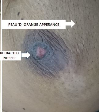
Most early breast cancer patients are asymptomatic and discovered during screening mammography. With increasing size, the patient may discover cancer as a lump that is felt accidentally, mostly during combing or showering. Breast pain is an unusual symptom that happens 5% of the time. The locally advanced disease may be presented with peau d'orange, frank ulceration, or fixation to the chest wall. Inflammatory breast cancer, an advanced form of breast cancer, frequently resembles breast abscess and presents with swelling, redness, and other local signs of inflammation. Paget disease of the nipple usually presents with nipple changes that must be differentiated from nipple eczema. 

Evaluation

Evaluation of Patients with breast cancer needs a triple assessment using clinical evaluation, imaging, and tissue biopsy. Mammography is the most commonly used modality for the diagnosis of breast cancer. Most of the asymptomatic cases are diagnosed during screening mammography. Breast cancer always presents as calcifications, dense lump, with or without architecture distortion. However, mammography is not sensitive in young women for whom breast ultrasonography can be used. Ultrasonography is useful in assessing the consistency and size of breast lumps. It has a great role in guided needle biopsy. Magnetic resonance imaging has good sensitivity for describing abnormalities in soft tissues, including the breast. It is indicated if there are occult lesions, or suspicion of multifocal or bilateral malignancy, especially ILC, and in the assessment of response to neoadjuvant chemotherapy, or when planning for breast conservation surgery and screening in the high-risk patient. Tissue biopsy is an important step in the evaluation of a breast cancer patient. There are different ways to take a tissue specimen, and these include fine-needle aspiration cytology, core biopsy (Trucut), and incisional or excisional biopsy.[12][13][14]

Treatment / Management

The 2 basic principles of treatment are to reduce the chance of local recurrence and the risk of metastatic spread. Surgery with or without radiotherapy achieves local control of cancer. When there is a risk for metastatic relapse, systemic therapy is indicated in the form of hormonal therapy, chemotherapy, targeted therapy, or any combination of these. In locally advanced disease, systemic therapy is used as a palliative therapy with a small or no role for surgery.[15][16][17]

Differential Diagnosis

  • Breast abscess
  • Fat necrosis
  • Fibroadenoma

Surgical Oncology

Surgery has a major role in the treatment of breast cancer. It is the basic way to use for local control of the disease. Radical mastectomy of Halsted, which removed the breast with axillary lymph node dissection and excision of both pectoralis muscles, is no longer recommended due to the high rate of morbidity without a survival benefit. Now, the modified radical mastectomy of Patey is more famous. It entails removing the whole breast tissue with a large part of the skin and the axillary lymph nodes. The pectoralis major and minor muscles are preserved. Breast-only removal without axillary dissection is referred to as simple mastectomy. This procedure can be performed in small tumors with negative sentinel lymph nodes. Breast-conserving surgery (BCS) is aimed at removing the tumor plus a rim of at least 1 cm of normal breast tissue (wide local excision). A quadrantectomy involves removing the entire segment of the breast that contains the tumor. The last 2 procedures are usually combined with axillary clearance through a separate incision. Axillary procedures may include sentinel lymph node biopsy, sampling, partial (II), or complete (III) axillary lymph node dissection. Lumpectomy is the removal of a benign mass without excision of the normal breast tissue.

Radiation Oncology

Radiation therapy has a significant role in local disease control. The risk of cancer recurrence decreases by about 50% at 10 years, and the risk of breast cancer death reduces by almost 20% at 15 years when radiation therapy follows BCS. However, radiation is not necessary for women 70 years of age and older with small, lymph node-negative, hormone receptor-positive (HR+) cancers because it has not been shown to improve survival in patients who take hormonal therapy for at least 5 years. Radiation therapy is beneficial in large tumors (greater to 5 cm) or if the tumor invades skin or chest wall and if there are positive lymph nodes. It can also be used as palliative therapy in advanced cases, such as a central nervous system (CNS) or bone metastasis. It can be delivered as external beam radiation, brachytherapy, or a combination of both.[18][19]

Medical Oncology

Chemotherapy, hormone therapy, and targeted therapy are the systemic therapies used in breast cancer management. A 25 percent reduction in the risk of relapse over a 10 to 15-year period using a first-generation chemotherapy regimen such as cyclophosphamide, methotrexate, and 5-fluorouracil (CMF) in a 6-month cycle. Anthracyclines (doxorubicin or epirubicin) and the newer agents such as the taxanes are modern regimens used for breast cancer. Three to 6-month period is used for adjuvant and neoadjuvant chemotherapy. Adjuvant treatment of early-stage HR+ breast cancer with tamoxifen for at least 5 years has been shown to reduce the recurrence rate by about half throughout the first 10 years and reduces breast cancer mortality by about 30% throughout the first 15 years.

More recently, studies have shown that extended use of adjuvant tamoxifen (10 years versus 5 years) further reduces the risk of breast cancer recurrence and mortality, so clinical practice guidelines now recommend consideration of adjuvant tamoxifen therapy for 10 years. The mainstay of treatment for most premenopausal women with HR+ tumors is tamoxifen. Some women may also benefit from surgical removal (oophorectomy) or chemical suppression of the ovaries, which are the main source of estrogen before menopause. Treatment guidelines recommend aromatase inhibitors (AIs) such as anastrozole should usually be included in the treatment of postmenopausal women with HR+ breast cancer. Targeted therapy is usually indicated in about 17% of breast cancers that overproduce the growth-promoting protein HER2/neu. Trastuzumab, the first approved drug, is a monoclonal antibody that directly targets the HER2 protein. It reduces the risk of recurrence and death by 52% and 33%, respectively, if combined with chemotherapy in HER2+ early breast cancer if compared to chemotherapy alone.[20][21]

Staging

Breast cancer staging is determined clinically by physical examination and imaging studies before treatment, and breast cancer stage is determined pathologically by pathologic examination of the primary tumor and regional lymph nodes after definitive surgical treatment. Staging is performed to group patients into risk categories that define prognosis and guide treatment recommendations for patients with a similar prognosis. Breast cancer is classified with the TNM classification system, which groups patients into 4 stage groupings based on the primary tumor size (T),  the regional lymph nodes status (N), and if there is any distant metastasis (M). The most widely used system is that of the American Joint Committee on Cancer:

Primary Tumor (T)  

Tis: Carcinoma in-situ, Paget‘s with no tumor

T1: Less than 2 cmT1a: 0.1 to 0.5 cmT1b: 0.5 to 1.0 cmT1c: 1.0 to 2.0 cm

T2: 2 to 5 cm

T3: Larger than 5 cm

T4T4a: Chest wall involvementT4b: Skin involvementT4c: Both 4a and 4bT4d: Inflammatory ca

Regional Lymph Nodes (N)

N1: Mobile ipsilateral axillary nodes

N2: Fixed/matted ipsilateral axillary nodes

N3N3a – Ipsilat infraclavicular nodesN3b – Ipsilat int mammary nodesN3c – Ipsilateral supraclavicular nodes

Distant Metastases (M)

M1: Distant metastases

Stage 0    Tis

Stage IT1N0

Stage IIT2N0, T3N0 T0N1, T1N1, T2N1

Stage III 

*skin, rib inv., matted LNs T3 N1 T0N2, T1N2, T2N2, T3N2 Any T, N3 T4, any N Locally advanced BC

Stage IV

M1 Adv. BC

Prognosis

The prognosis of early breast cancer is quietly good. Stage 0 and stage I both have a 100% 5-year survival rate. The 5-year survival rate of stage II and stage III breast cancer is about 93% and 72%, respectively. When the disease spreads systemically, its prognosis worsens dramatically. Only 22% of stage IV breast cancer patients will survive their next 5 years.

Complications

Complications can arise from the treatment, whether chemotherapy, radiation, hormonal therapy, or surgery.

Surgical complications include:

  • Infection
  • Pain
  • Bleeding
  • Cosmetic issues
  • Permanent scarring
  • Alteration or loss of sensation in the chest area and reconstructed breasts

Chemotherapy complications include:

  • Nausea/vomiting and diarrhea
  • Hair loss
  • Memory loss ("chemo brain")
  • Vaginal dryness
  • Menopausal symptoms/fertility issues
  • Neuropathy

Complications accompanying hormonal therapy include:

  • Hot flashes
  • Vaginal discharge dryness
  • Fatigue
  • Nausea
  • Impotence in males with breast cancer

Radiation can result in the following complications:

  • Pain and skin changes
  • Fatigue
  • Nausea
  • Hair loss
  • Heart and lung issues (long-term)
  • Neuropathy

Deterrence and Patient Education

Patients usually require counseling to deal with the condition and treatment. They should be put in touch with psychological counseling, and there are also support groups available.

Pearls and Other Issues

Breast cancer patients are advised to be followed up for life to detect early recurrence and spread. Yearly or biannual follow-up mammography is recommended for the treated and the other breast. The patient must be informed that they must visit a breast clinic if they have any suspicious manifestations. Currently, there is no role for repeated measurements of tumor markers or doing follow-up imaging other than mammography.

Enhancing Healthcare Team Outcomes

After the treatment of breast cancer, long-term follow-up is necessary. There is a risk of local and distant relapse, and hence an interprofessional team approach is necessary. The women need regular mammograms and a pelvic exam. Also, women with risk factors for osteoporosis need a bone density exam and monitoring for tumor markers for metastatic disease. For those who are about to undergo radiation therapy, a baseline echo and cardiac evaluation are necessary. Even though many types of integrative therapies have been developed to help women with breast cancer, evidence for the majority of these treatments is weak or lacking.[22]

Outcomes

Over the past four decades, the survival rates of most breast cancer patients have improved. Of note is that the presence of breast cancer has gradually slowed down over the past decade, which may be due to earlier detection and improved treatments. The prognosis for patients with breast cancer is highly dependent on the status of axillary lymph nodes. The higher the number of positive lymph nodes, the worse the outcome. In general, hormone-responsive tumors tend to have a better outcome. In breast cancer survivors, adverse cardiac events are common; this is partly due to the cardiotoxic drugs to treat cancer and the presence of traditional risk factors for heart disease. The onus is on the healthcare provider to reduce the modifiable risk factors and lower the risk of adverse cardiac events.[1][23] [Level 5)


(Click Image to Enlarge)
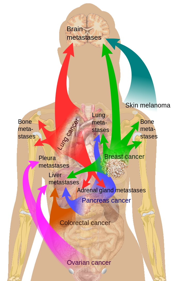
Main sites of metastases for common cancer types, Brain, Skin Melanoma, Bone Metastases, Lung Metastases, Breast Cancer, Adre
Main sites of metastases for common cancer types, Brain, Skin Melanoma, Bone Metastases, Lung Metastases, Breast Cancer, Adrenal gland metastases, Pancreas cancer, Colorectal cancer, Ovarian cancer
Contributed by Wikimedia Commons,"Medical gallery of Mikael Häggström 2014" (Public Domain)

(Click Image to Enlarge)
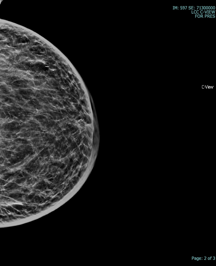
CC mammographic view of the left breast demonstrates skin thickening, trabecular coasrening/diffusely increased breast density and malignant type calcifications in this patient with biopsy proven inflammatory breast cancer
CC mammographic view of the left breast demonstrates skin thickening, trabecular coasrening/diffusely increased breast density and malignant type calcifications in this patient with biopsy proven inflammatory breast cancer.
Contributed by Hassana Barazi, MD

(Click Image to Enlarge)
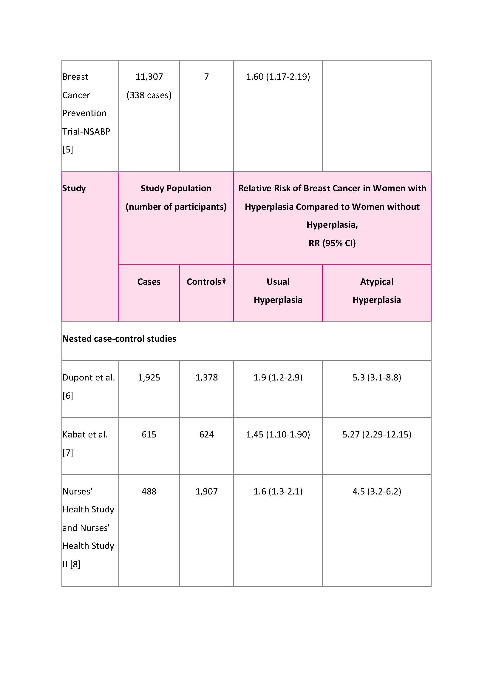
Table 1.b Risk breast cancer consortium surveillance
5. Kerlikowske K, Gard CC, Tice JA, et al. for the Breast Cancer Surveillance Consortium. Risk factors that increase risk of estrogen receptor-positive and -negative breast cancer. J Natl Cancer Inst. 109(5): djw276, 2016.

(Click Image to Enlarge)
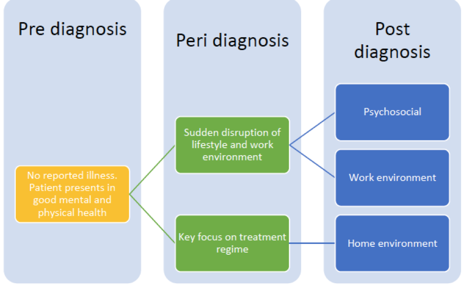
Interprofessional care diagram Breast cancer patients
Contributed by Kathryn Malherbe (PhD candidate)

(Click Image to Enlarge)
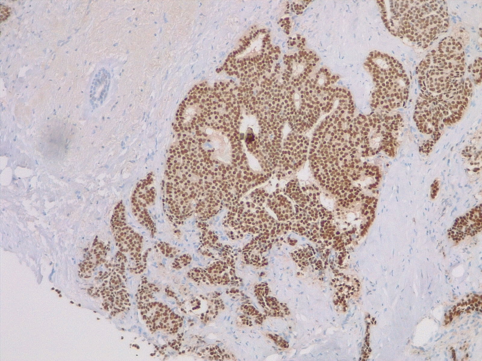
Estrogen receptor expression in breast carcinoma, ER stain 4x
Contributed by Fabiola Farci, MD

(Click Image to Enlarge)
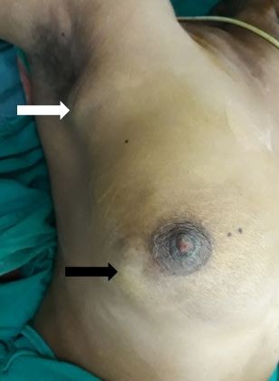
Ca breast with axillary lymphadenopathy
Contributed by Sunil Munakomi, MD

(Click Image to Enlarge)
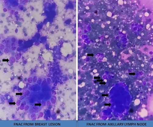
FNAC from breast cancer
Contributed by Sunil Munakomi, MD

(Click Image to Enlarge)
Clinical findings in carcinoma breast
Contributed by Sunil Munakomi, MD
Details

Editor:

Troy Ferguson

Updated:

9/26/2022 7:30:23 PM

References


[1]

Mahvi DA, Liu R, Grinstaff MW, Colson YL, Raut CP. Local Cancer Recurrence: The Realities, Challenges, and Opportunities for New Therapies. CA: a cancer journal for clinicians. 2018 Nov:68(6):488-505. doi: 10.3322/caac.21498. Epub 2018 Oct 17     [PubMed PMID: 30328620]


[2]

Narod SA, Personalised medicine and population health: breast and ovarian cancer. Human genetics. 2018 Oct     [PubMed PMID: 30328515]


[3]

Cain EH, Saha A, Harowicz MR, Marks JR, Marcom PK, Mazurowski MA. Multivariate machine learning models for prediction of pathologic response to neoadjuvant therapy in breast cancer using MRI features: a study using an independent validation set. Breast cancer research and treatment. 2019 Jan:173(2):455-463. doi: 10.1007/s10549-018-4990-9. Epub 2018 Oct 16     [PubMed PMID: 30328048]

Level 1 (high-level) evidence

[4]

PDQ Screening and Prevention Editorial Board. Breast Cancer Screening (PDQ®): Health Professional Version. PDQ Cancer Information Summaries. 2002:():     [PubMed PMID: 26389344]


[5]

Doren A, Vecchiola A, Aguirre B, Villaseca P. Gynecological-endocrinological aspects in women carriers of BRCA1/2 gene mutations. Climacteric : the journal of the International Menopause Society. 2018 Dec:21(6):529-535. doi: 10.1080/13697137.2018.1514006. Epub 2018 Oct 8     [PubMed PMID: 30295091]

Level 2 (mid-level) evidence

[6]

Parada H Jr, Sun X, Tse CK, Olshan AF, Troester MA. Lifestyle Patterns and Survival Following Breast Cancer in the Carolina Breast Cancer Study. Epidemiology (Cambridge, Mass.). 2019 Jan:30(1):83-92. doi: 10.1097/EDE.0000000000000933. Epub     [PubMed PMID: 30299404]


[7]

White AJ, Bradshaw PT, Hamra GB. Air pollution and Breast Cancer: A Review. Current epidemiology reports. 2018 Jun:5(2):92-100. doi: 10.1007/s40471-018-0143-2. Epub 2018 Mar 27     [PubMed PMID: 30271702]


[8]

Gucalp A, Traina TA, Eisner JR, Parker JS, Selitsky SR, Park BH, Elias AD, Baskin-Bey ES, Cardoso F. Male breast cancer: a disease distinct from female breast cancer. Breast cancer research and treatment. 2019 Jan:173(1):37-48. doi: 10.1007/s10549-018-4921-9. Epub 2018 Sep 28     [PubMed PMID: 30267249]


[9]

Clark BZ, Onisko A, Assylbekova B, Li X, Bhargava R, Dabbs DJ. Breast cancer global tumor biomarkers: a quality assurance study of intratumoral heterogeneity. Modern pathology : an official journal of the United States and Canadian Academy of Pathology, Inc. 2019 Mar:32(3):354-366. doi: 10.1038/s41379-018-0153-0. Epub 2018 Oct 16     [PubMed PMID: 30327501]

Level 2 (mid-level) evidence

[10]

Liedtke C, Kolberg HC, Kerschke L, Görlich D, Bauerfeind I, Fehm T, Fleige B, Helms G, Lebeau A, Stäbler A, Schmatloch S, Hausschild M, Schwentner L, von Minckwitz G, Loibl S, Untch M, Kühn T. Systematic analysis of parameters predicting pathological axillary status (ypN0 vs. ypN+) in patients with breast cancer converting from cN+ to ycN0 through primary systemic therapy (PST). Clinical & experimental metastasis. 2018 Dec:35(8):777-783. doi: 10.1007/s10585-018-9938-2. Epub 2018 Oct 15     [PubMed PMID: 30324492]

Level 1 (high-level) evidence

[11]

Kitamura M, Nakayama T, Mukaisho KI, Mori T, Umeda T, Moritani S, Kushima R, Tani M, Sugihara H. Progression Potential of Ductal Carcinoma in situ Assessed by Genomic Copy Number Profiling. Pathobiology : journal of immunopathology, molecular and cellular biology. 2019:86(2-3):92-101. doi: 10.1159/000492833. Epub 2018 Oct 17     [PubMed PMID: 30332671]


[12]

Radovic N, Ivanac G, Divjak E, Biondic I, Bulum A, Brkljacic B. Evaluation of Breast Cancer Morphology Using Diffusion-Weighted and Dynamic Contrast-Enhanced MRI: Intermethod and Interobserver Agreement. Journal of magnetic resonance imaging : JMRI. 2019 May:49(5):1381-1390. doi: 10.1002/jmri.26332. Epub 2018 Oct 16     [PubMed PMID: 30325549]


[13]

Pediconi F, Marzocca F, Cavallo Marincola B, Napoli A. MRI-guided treatment in the breast. Journal of magnetic resonance imaging : JMRI. 2018 Dec:48(6):1479-1488. doi: 10.1002/jmri.26282. Epub 2018 Oct 14     [PubMed PMID: 30318672]


[14]

Watanabe Y, Anan K. The decision to perform or omit sentinel lymph node biopsy during mastectomy for ductal carcinoma in situ should be tailored in accordance with preoperative findings. Breast cancer (Tokyo, Japan). 2019 Mar:26(2):261-262. doi: 10.1007/s12282-018-0917-x. Epub 2018 Oct 16     [PubMed PMID: 30328007]


[15]

Rocque GB, Williams CP, Kenzik KM, Jackson BE, Azuero A, Halilova KI, Ingram SA, Pisu M, Forero A, Bhatia S. Concordance with NCCN treatment guidelines: Relations with health care utilization, cost, and mortality in breast cancer patients with secondary metastasis. Cancer. 2018 Nov 1:124(21):4231-4240. doi: 10.1002/cncr.31694. Epub 2018 Oct 14     [PubMed PMID: 30317547]


[16]

Vande Perre P, Toledano D, Corsini C, Escriba E, Laporte M, Bertet H, Yauy K, Toledano A, Galibert V, Baudry K, Clotet L, Million E, Picot MC, Geneviève D, Pujol P. Role of the general practitioner in the care of BRCA1 and BRCA2 mutation carriers: General practitioner and patient perspectives. Molecular genetics & genomic medicine. 2018 Nov:6(6):957-965. doi: 10.1002/mgg3.464. Epub 2018 Oct 11     [PubMed PMID: 30308700]

Level 3 (low-level) evidence

[17]

Seroussi B, Lamy JB, Muro N, Larburu N, Sekar BD, Guézennec G, Bouaud J. Implementing Guideline-Based, Experience-Based, and Case-Based Approaches to Enrich Decision Support for the Management of Breast Cancer Patients in the DESIREE Project. Studies in health technology and informatics. 2018:255():190-194     [PubMed PMID: 30306934]

Level 3 (low-level) evidence

[18]

Wang X, Xu L, Yin Z, Wang D, Wang Q, Xu K, Zhao J, Zhao L, Yuan Z, Wang P. Locoregional recurrence-associated factors and risk-adapted postmastectomy radiotherapy for breast cancer staged in cT1-2N0-1 after neoadjuvant chemotherapy. Cancer management and research. 2018:10():4105-4112. doi: 10.2147/CMAR.S173628. Epub 2018 Oct 2     [PubMed PMID: 30323666]


[19]

Tang L, Matsushita H, Jingu K. Controversial issues in radiotherapy after breast-conserving surgery for early breast cancer in older patients: a systematic review. Journal of radiation research. 2018 Nov 1:59(6):789-793. doi: 10.1093/jrr/rry071. Epub     [PubMed PMID: 30321392]

Level 1 (high-level) evidence

[20]

Wu YT, Xu Z, Zhang K, Wu JS, Li X, Arshad B, Li YC, Wang ZL, Li HY, Wu KN, Kong LQ. Efficacy and cardiac safety of the concurrent use of trastuzumab and anthracycline-based neoadjuvant chemotherapy for HER2-positive breast cancer: a systematic review and meta-analysis. Therapeutics and clinical risk management. 2018:14():1789-1797. doi: 10.2147/TCRM.S176214. Epub 2018 Sep 26     [PubMed PMID: 30310287]

Level 1 (high-level) evidence

[21]

Dieci MV, Vernaci G, Guarneri V. Escalation and de-escalation in HER2 positive early breast cancer. Current opinion in oncology. 2019 Jan:31(1):35-42. doi: 10.1097/CCO.0000000000000492. Epub     [PubMed PMID: 30325338]

Level 3 (low-level) evidence

[22]

Hodis HN, Sarrel PM. Menopausal hormone therapy and breast cancer: what is the evidence from randomized trials? Climacteric : the journal of the International Menopause Society. 2018 Dec:21(6):521-528. doi: 10.1080/13697137.2018.1514008. Epub 2018 Oct 9     [PubMed PMID: 30296850]

Level 1 (high-level) evidence

[23]

Gautam S, Sylwestrzak G, Barron J, Chen X, Eleff M, Debono D, Nguyen A, Fisch M. Results From a Health Insurer's Clinical Pathway Program in Breast Cancer. Journal of oncology practice. 2018 Oct 15:():JOP1800157. doi: 10.1200/JOP.18.00157. Epub 2018 Oct 15     [PubMed PMID: 30321101]


[24]

Wapnir IL, Dignam JJ, Fisher B, Mamounas EP, Anderson SJ, Julian TB, Land SR, Margolese RG, Swain SM, Costantino JP, Wolmark N. Long-term outcomes of invasive ipsilateral breast tumor recurrences after lumpectomy in NSABP B-17 and B-24 randomized clinical trials for DCIS. Journal of the National Cancer Institute. 2011 Mar 16:103(6):478-88. doi: 10.1093/jnci/djr027. Epub 2011 Mar 11     [PubMed PMID: 21398619]

Level 1 (high-level) evidence

[25]

Litière S, Werutsky G, Fentiman IS, Rutgers E, Christiaens MR, Van Limbergen E, Baaijens MH, Bogaerts J, Bartelink H. Breast conserving therapy versus mastectomy for stage I-II breast cancer: 20 year follow-up of the EORTC 10801 phase 3 randomised trial. The Lancet. Oncology. 2012 Apr:13(4):412-9. doi: 10.1016/S1470-2045(12)70042-6. Epub 2012 Feb 27     [PubMed PMID: 22373563]

Level 1 (high-level) evidence

[26]

Kirby AM. Updated ASTRO guidelines on accelerated partial breast irradiation (APBI): to whom can we offer APBI outside a clinical trial? The British journal of radiology. 2018 May:91(1085):20170565. doi: 10.1259/bjr.20170565. Epub 2018 Mar 23     [PubMed PMID: 29513031]


[27]

Vicini FA, Cecchini RS, White JR, Arthur DW, Julian TB, Rabinovitch RA, Kuske RR, Ganz PA, Parda DS, Scheier MF, Winter KA, Paik S, Kuerer HM, Vallow LA, Pierce LJ, Mamounas EP, McCormick B, Costantino JP, Bear HD, Germain I, Gustafson G, Grossheim L, Petersen IA, Hudes RS, Curran WJ Jr, Bryant JL, Wolmark N. Long-term primary results of accelerated partial breast irradiation after breast-conserving surgery for early-stage breast cancer: a randomised, phase 3, equivalence trial. Lancet (London, England). 2019 Dec 14:394(10215):2155-2164. doi: 10.1016/S0140-6736(19)32514-0. Epub 2019 Dec 5     [PubMed PMID: 31813636]

Level 1 (high-level) evidence

[28]

Bartelink H, Horiot JC, Poortmans PM, Struikmans H, Van den Bogaert W, Fourquet A, Jager JJ, Hoogenraad WJ, Oei SB, Wárlám-Rodenhuis CC, Pierart M, Collette L. Impact of a higher radiation dose on local control and survival in breast-conserving therapy of early breast cancer: 10-year results of the randomized boost versus no boost EORTC 22881-10882 trial. Journal of clinical oncology : official journal of the American Society of Clinical Oncology. 2007 Aug 1:25(22):3259-65     [PubMed PMID: 17577015]

Level 1 (high-level) evidence

[29]

Overgaard M, Nielsen HM, Tramm T, Højris I, Grantzau TL, Alsner J, Offersen BV, Overgaard J, DBCG Radiotherapy Group. Postmastectomy radiotherapy in high-risk breast cancer patients given adjuvant systemic therapy. A 30-year long-term report from the Danish breast cancer cooperative group DBCG 82bc trial. Radiotherapy and oncology : journal of the European Society for Therapeutic Radiology and Oncology. 2022 May:170():4-13. doi: 10.1016/j.radonc.2022.03.008. Epub 2022 Mar 11     [PubMed PMID: 35288227]


[30]

Donker M, van Tienhoven G, Straver ME, Meijnen P, van de Velde CJ, Mansel RE, Cataliotti L, Westenberg AH, Klinkenbijl JH, Orzalesi L, Bouma WH, van der Mijle HC, Nieuwenhuijzen GA, Veltkamp SC, Slaets L, Duez NJ, de Graaf PW, van Dalen T, Marinelli A, Rijna H, Snoj M, Bundred NJ, Merkus JW, Belkacemi Y, Petignat P, Schinagl DA, Coens C, Messina CG, Bogaerts J, Rutgers EJ. Radiotherapy or surgery of the axilla after a positive sentinel node in breast cancer (EORTC 10981-22023 AMAROS): a randomised, multicentre, open-label, phase 3 non-inferiority trial. The Lancet. Oncology. 2014 Nov:15(12):1303-10. doi: 10.1016/S1470-2045(14)70460-7. Epub 2014 Oct 15     [PubMed PMID: 25439688]

Level 1 (high-level) evidence

[31]

Whelan TJ, Olivotto IA, Parulekar WR, Ackerman I, Chua BH, Nabid A, Vallis KA, White JR, Rousseau P, Fortin A, Pierce LJ, Manchul L, Chafe S, Nolan MC, Craighead P, Bowen J, McCready DR, Pritchard KI, Gelmon K, Murray Y, Chapman JA, Chen BE, Levine MN, MA.20 Study Investigators. Regional Nodal Irradiation in Early-Stage Breast Cancer. The New England journal of medicine. 2015 Jul 23:373(4):307-16. doi: 10.1056/NEJMoa1415340. Epub     [PubMed PMID: 26200977]


[32]

Choi KH, Ahn SJ, Jeong JU, Yu M, Kim JH, Jeong BK, Lee JH, Kim SH, Lee JH. Postoperative radiotherapy with intensity-modulated radiation therapy versus 3-dimensional conformal radiotherapy in early breast cancer: A randomized clinical trial of KROG 15-03. Radiotherapy and oncology : journal of the European Society for Therapeutic Radiology and Oncology. 2021 Jan:154():179-186. doi: 10.1016/j.radonc.2020.09.043. Epub 2020 Sep 24     [PubMed PMID: 32980384]

Level 1 (high-level) evidence

[33]

Pignol JP, Truong P, Rakovitch E, Sattler MG, Whelan TJ, Olivotto IA. Ten years results of the Canadian breast intensity modulated radiation therapy (IMRT) randomized controlled trial. Radiotherapy and oncology : journal of the European Society for Therapeutic Radiology and Oncology. 2016 Dec:121(3):414-419. doi: 10.1016/j.radonc.2016.08.021. Epub 2016 Sep 13     [PubMed PMID: 27637858]

Level 1 (high-level) evidence

[34]

Darby SC, Ewertz M, McGale P, Bennet AM, Blom-Goldman U, Brønnum D, Correa C, Cutter D, Gagliardi G, Gigante B, Jensen MB, Nisbet A, Peto R, Rahimi K, Taylor C, Hall P. Risk of ischemic heart disease in women after radiotherapy for breast cancer. The New England journal of medicine. 2013 Mar 14:368(11):987-98. doi: 10.1056/NEJMoa1209825. Epub     [PubMed PMID: 23484825]


[35]

Epler GR, Kelly EM. Systematic review of postradiotherapy bronchiolitis obliterans organizing pneumonia in women with breast cancer. The oncologist. 2014 Dec:19(12):1216-26. doi: 10.1634/theoncologist.2014-0041. Epub 2014 Oct 31     [PubMed PMID: 25361622]

Level 1 (high-level) evidence

[36]

Taghian AG, Assaad SI, Niemierko A, Floyd SR, Powell SN. Is a reduction in radiation lung volume and dose necessary with paclitaxel chemotherapy for node-positive breast cancer? International journal of radiation oncology, biology, physics. 2005 Jun 1:62(2):386-91     [PubMed PMID: 15890579]


[37]

Williams NR, Williams S, Kanapathy M, Naderi N, Vavourakis V, Mosahebi A. Radiation-induced fibrosis in breast cancer: A protocol for an observational cross-sectional pilot study for personalised risk estimation and objective assessment. International journal of surgery protocols. 2019:14():9-13. doi: 10.1016/j.isjp.2019.02.002. Epub 2019 Feb 13     [PubMed PMID: 31851743]

Level 2 (mid-level) evidence

[38]

Collette S, Collette L, Budiharto T, Horiot JC, Poortmans PM, Struikmans H, Van den Bogaert W, Fourquet A, Jager JJ, Hoogenraad W, Mueller RP, Kurtz J, Morgan DA, Dubois JB, Salamon E, Mirimanoff R, Bolla M, Van der Hulst M, Wárlám-Rodenhuis CC, Bartelink H, EORTC Radiation Oncology Group. Predictors of the risk of fibrosis at 10 years after breast conserving therapy for early breast cancer: a study based on the EORTC Trial 22881-10882 'boost versus no boost'. European journal of cancer (Oxford, England : 1990). 2008 Nov:44(17):2587-99. doi: 10.1016/j.ejca.2008.07.032. Epub 2008 Aug 29     [PubMed PMID: 18757193]


[39]

Jacobson G, Bhatia S, Smith BJ, Button AM, Bodeker K, Buatti J. Randomized trial of pentoxifylline and vitamin E vs standard follow-up after breast irradiation to prevent breast fibrosis, evaluated by tissue compliance meter. International journal of radiation oncology, biology, physics. 2013 Mar 1:85(3):604-8. doi: 10.1016/j.ijrobp.2012.06.042. Epub 2012 Jul 28     [PubMed PMID: 22846413]

Level 1 (high-level) evidence

[40]

Gross JP, Whelan TJ, Parulekar WR, Chen BE, Rademaker AW, Helenowski IB, Donnelly ED, Strauss JB. Development and Validation of a Nomogram to Predict Lymphedema After Axillary Surgery and Radiation Therapy in Women With Breast Cancer From the NCIC CTG MA.20 Randomized Trial. International journal of radiation oncology, biology, physics. 2019 Sep 1:105(1):165-173. doi: 10.1016/j.ijrobp.2019.05.002. Epub 2019 May 11     [PubMed PMID: 31085285]

Level 1 (high-level) evidence

[41]

DiSipio T, Rye S, Newman B, Hayes S. Incidence of unilateral arm lymphoedema after breast cancer: a systematic review and meta-analysis. The Lancet. Oncology. 2013 May:14(6):500-15. doi: 10.1016/S1470-2045(13)70076-7. Epub 2013 Mar 27     [PubMed PMID: 23540561]

Level 1 (high-level) evidence

[42]

Bartels SAL, Donker M, Poncet C, Sauvé N, Straver ME, van de Velde CJH, Mansel RE, Blanken C, Orzalesi L, Klinkenbijl JHG, van der Mijle HCJ, Nieuwenhuijzen GAP, Veltkamp SC, van Dalen T, Marinelli A, Rijna H, Snoj M, Bundred NJ, Merkus JWS, Belkacemi Y, Petignat P, Schinagl DAX, Coens C, van Tienhoven G, van Duijnhoven F, Rutgers EJT. Radiotherapy or Surgery of the Axilla After a Positive Sentinel Node in Breast Cancer: 10-Year Results of the Randomized Controlled EORTC 10981-22023 AMAROS Trial. Journal of clinical oncology : official journal of the American Society of Clinical Oncology. 2023 Apr 20:41(12):2159-2165. doi: 10.1200/JCO.22.01565. Epub 2022 Nov 16     [PubMed PMID: 36383926]

Level 1 (high-level) evidence

[43]

Pierce SM, Recht A, Lingos TI, Abner A, Vicini F, Silver B, Herzog A, Harris JR. Long-term radiation complications following conservative surgery (CS) and radiation therapy (RT) in patients with early stage breast cancer. International journal of radiation oncology, biology, physics. 1992:23(5):915-23     [PubMed PMID: 1639653]


[44]

Meric F, Buchholz TA, Mirza NQ, Vlastos G, Ames FC, Ross MI, Pollock RE, Singletary SE, Feig BW, Kuerer HM, Newman LA, Perkins GH, Strom EA, McNeese MD, Hortobagyi GN, Hunt KK. Long-term complications associated with breast-conservation surgery and radiotherapy. Annals of surgical oncology. 2002 Jul:9(6):543-9     [PubMed PMID: 12095969]


[45]

Grantzau T, Overgaard J. Risk of second non-breast cancer after radiotherapy for breast cancer: a systematic review and meta-analysis of 762,468 patients. Radiotherapy and oncology : journal of the European Society for Therapeutic Radiology and Oncology. 2015 Jan:114(1):56-65. doi: 10.1016/j.radonc.2014.10.004. Epub 2014 Nov 7     [PubMed PMID: 25454172]

Level 1 (high-level) evidence

[46]

Ye JC, Yan W, Christos P, Nori D, Chao KS, Ravi A. Second cancer, breast cancer, and cardiac mortality in stage T1aN0 breast cancer patients with or without external beam radiation therapy: a national registry study. Clinical breast cancer. 2015 Feb:15(1):54-9. doi: 10.1016/j.clbc.2014.07.003. Epub 2014 Aug 15     [PubMed PMID: 25223278]


[47]

Dracham CB, Shankar A, Madan R. Radiation induced secondary malignancies: a review article. Radiation oncology journal. 2018 Jun:36(2):85-94. doi: 10.3857/roj.2018.00290. Epub 2018 Jun 29     [PubMed PMID: 29983028]