Continuing Education Activity
Alzheimer disease (AD) is the most common cause of a decline in cognitive ability. It is a neurodegenerative disorder that usually affects people over the age of 65 with the involvement of language, memory, comprehension, attention, judgment and reasoning. This activity reviews the evaluation and management of Alzheimer disease and explains the role of the interprofessional team in improving care for patients with this condition.
Objectives:
- Identify the risk factors associated with Alzheimer disease.
- Describe the atypical presentations of Alzheimer disease.
- Summarize and differentiate between the preclinical, mild, moderate and severe stages of Alzheimer disease.
- Outline the importance of collaboration and communication among the interprofessional team to enhance delivery of care for patients affected by Alzheimer disease.
Introduction
Dementia is a general term that refers to a decline in cognitive ability severe enough to interfere with activities of daily living. Alzheimer's disease (AD) is the most common type of dementia, accounting for at least two-thirds of cases of dementia in people age 65 and older. Alzheimer's disease is a neurodegenerative disease with insidious onset and progressive impairment of behavioral and cognitive functions including memory, comprehension, language, attention, reasoning, and judgment. It is the sixth leading cause of death in the United States. Onset before 65 years of age (early onset) is unusual and seen in less than 10% of Alzheimer's disease patients. There is no cure for Alzheimer's disease, although there are treatments available that may improve some symptoms.
Symptoms of Alzheimer's disease depend on the stage of the disease. Alzheimer's disease is classified into preclinical or presymptomatic, mild, and dementia-stage depending on the degree of cognitive impairment. These stages are different from the DSM-5 classification of Alzheimer's disease. The initial and most common presenting symptom is episodic short-term memory loss with relative sparing of long-term memory and can be elicited in most patients even when not the presenting symptom. Short-term memory impairment is followed by impairment in problem-solving, judgment, executive functioning, lack of motivation and disorganization, leading to problems with multitasking and abstract thinking. In the early stages, impairment in executive functioning ranges from subtle to significant. This is followed by language disorder and impairment of visuospatial skills. Neuropsychiatric symptoms like apathy, social withdrawal, disinhibition, agitation, psychosis, and wandering are also common in the mid to late stages. Difficulty performing learned motor tasks (dyspraxia), olfactory dysfunction, sleep disturbances, extrapyramidal motor signs like dystonia, akathisia, and parkinsonian symptoms occur late in the disease. This is followed by primitive reflexes, incontinence, and total dependence on caregivers.[1],[2],[3]
Etiology
Alzheimer's disease is a gradual and progressive neurodegenerative disease caused by neuronal cell death. It typically starts in the entorhinal cortex in the hippocampus. There is a genetic role identified for both early and late-onset Alzheimer's disease. Trisomy 21 is a risk factor for early-onset dementia. Several risk factors have been associated with Alzheimer's disease. Increasing age is the most important risk factor for Alzheimer's disease. Traumatic head injury, depression, cardiovascular and cerebrovascular disease, higher parental age, smoking, family history of dementia, increased homocysteine levels and presence of APOE e4 allele are known to increase the risk of Alzheimer's disease. Higher education, use of estrogen by women, use of anti-inflammatory agents, leisure activities like reading or playing musical instruments, healthy diet and regular aerobic exercise is known to decrease the risk of Alzheimer's disease. Having a first-degree relative with Alzheimer's disease increases the risk of developing Alzheimer's disease by 10% to 30%. Individuals with 2 or more siblings with late-onset Alzheimer disease increase their risk of getting Alzheimer's disease by 3-fold as compared to the general population.[4],[5],[6]
Epidemiology
Alzheimer's disease is typically a disease of old age. The global prevalence of dementia is reported to be as high as 24 million and is predicted to increase 4 times by the year 2050. The estimated health care cost of Alzheimer's disease is $172 billion per year in the United States alone. In 2011, the United States had an estimated 4.5 million people age sixty-five and above, living with clinical Alzheimer's disease. The incidence of Alzheimer's disease doubles every 5 years, after the age of 65. Age-specific incidence increases significantly from less than 1% per year before 65 years of age to 6% per year after 85 years of age. Prevalence rates increase from 10% after the age of 65 to 40 % after the age of 85. Incidence rates of Alzheimer's disease are slightly higher for women, especially after 85 years of age.
Pathophysiology
Alzheimer's disease is characterized by an accumulation of abnormal neuritic plaques and neurofibrillary tangles.
Plaques are spherical microscopic lesions that have a core of extracellular amyloid beta-peptide surrounded by enlarged axonal endings. Beta-amyloid peptide is derived from a transmembrane protein known as an amyloid precursor protein (APP). The beta-amyloid peptide is cleaved from APP by the action of proteases named alpha, beta, and gamma-secretase. Usually, APP is cleaved by either alpha or beta-secretase and the tiny fragments formed by them are not toxic to neurons. However, sequential cleavage by beta and then gamma-secretase results in 42 amino acid peptides (beta-amyloid 42). Elevation in levels of beta-amyloid 42 leads to aggregation of amyloid that causes neuronal toxicity. Beta-amyloid 42 favors the formation of aggregated fibrillary amyloid protein over normal APP degradation. APP gene is located on chromosome 21, one of the regions linked to familial Alzheimer's disease. Amyloid deposition occurs around meningeal and cerebral vessels and gray matter in Alzheimer's disease. Gray matter deposits are multifocal and coalesce to form milliary structures called plaques. However, brain scans have noted amyloid plaques in some persons without dementia and then other persons had dementia but brain scans did not find any plaques.
Neurofibrillary tangles are fibrillary intracytoplasmic structures in neurons formed by a protein called tau. The primary function of the tau protein is to stabilize axonal microtubules. Microtubules run along neuronal axons and are essential for intracellular transport. Microtubule assembly is held together by tau protein. In Alzheimer's disease, due to aggregation of extracellular beta-amyloid, there is hyperphosphorylation of tau which then causes the formation of tau aggregates. Tau aggregates form twisted paired helical filaments known as neurofibrillary tangles. They occur first in the hippocampus and then may be seen throughout the cerebral cortex. Tau-aggregates are deposited within the neurons. There is a staging system developed by Braak and Braak based on the topographical staging of neurofibrillary tangles into 6 stages, and this Braak staging is an integral part of the National Institute on Aging and Reagan Institute neuropathological criteria for the diagnosis of Alzheimer disease. Tangles are more strongly correlated to Alzheimer's than the plaques are.
Another feature of Alzheimer's disease is granulovacuolar degeneration of hippocampal pyramidal cells by amyloid angiopathy. Some reports indicate that cognitive decline correlates more with a decrease in the density of presynaptic boutons from pyramidal neurons in laminae III and IV, rather than an increase in the number of plaques.
Neuronal loss in Nucleus Basalis of Myenert, leading to low Acetylcholine has also been noted.
Vascular contribution to the neurodegenerative process of Alzheimer's disease is not fully determined. The risk of dementia is increased fourfold with subcortical infarcts. The cerebrovascular disease also exaggerates the degree of dementia and its rate of progression.[7],[8],[9]
Genetic Basis of Alzheimer Disease
Alzheimer's disease can be inherited as an autosomal dominant disorder with nearly complete penetrance. The autosomal dominant form of the disease is linked to mutations in 3 genes: AAP gene on chromosome 21, Presenilin1 (PSEN1) on chromosome 14, and Presenilin 2 (PSEN2) on chromosome 1. APP mutations may lead to increased generation and aggregation of beta-amyloid peptide. PSEN1 and PSEN2 mutations lead to aggregation of beta-amyloid by interfering with the processing of gamma-secretase. Mutations in these 3 genes account for about 5 % to 10 % of all the cases and about the majority of early-onset Alzheimer's disease.
Apolipoprotein E is a regulator of lipid metabolism that has an affinity for beta-amyloid protein and is another genetic marker that increases the risk of Alzheimer's disease. Isoform e4 of APOE gene (located on chromosome 19) has been associated with more sporadic and familial forms of Alzheimer's disease that present after age 65. The presence of one APOEe4 allele does not always lead to Alzheimer's disease, but among persons carrying one APOE- e4 allele about 50% have Alzheimer's disease and those having two alleles, 90% develop Alzheimer disease. Each APOE e4 allele also lowers the age of disease onset. The presence of the APOE e4 allele is an important risk factor for Alzheimer's disease.
Variants in the gene for the sortilin receptor, SORT1, which is essential for transporting APP from cell surface to Golgi-endoplasmic reticulum complex, have been found in familial and sporadic forms of Alzheimer disease.[4]
History and Physical
A good history and physical examination are the keys to diagnosis. It is also essential to take a history from the family and caregivers as some patients may lack insight into their disease. It is vital to characterize onset and early symptoms to differentiate from other types of dementia. It is important to obtain a good assessment of functional abilities like basic and individual activities of daily living.
A complete physical examination with a detailed neurological exam and mental status examination is needed to evaluate the disease stage and rule out other conditions. Comprehensive clinical assessment can provide reasonable diagnostic accuracy in most patients. A detailed neurological examination is essential to rule out other conditions. In Alzheimer's disease, the neurological exam is usually normal. The physical exam is normal except for anosmia. Anosmia is also found in patients with Parkinson's disease, dementia with Lewy bodies, and TBI with or without dementia, but not in those with VCI or depression. In the advanced stages of Alzheimer's disease, patients do not have lateralized signs. They eventually become mute, fail to respond to verbal requests, remain confined to bed, and frequently slip into a persistent vegetative state. A mental status examination should assess concentration, attention, recent and remote memory, language, visuospatial functioning, praxis, and executive functioning.
Brief standard examinations like the mini-mental status examination are less sensitive and specific, although they can be used for screening.
All follow-up visits should include a full mental status examination to evaluate disease progression and the development of neuropsychiatric symptoms.
Evaluation
Routine laboratory tests show no specific abnormality. Complete blood count (CBC), complete metabolic panel (CMP), thyroid-stimulating hormone (TSH), B12 are usually checked to rule out other causes.[10],[11],[12]
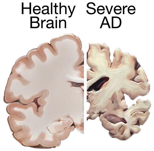
CT brain shows cerebral atrophy and widened the third ventricle. It is suggestive but it's nonspecific because these abnormalities are also present in other illnesses and people with normal age-related changes.
Cerebrospinal fluid (CSF) analysis for low beta-amyloid 42 and elevated tau is helpful in the diagnosis of the preclinical stage.
EEG typically shows a generalized slowing with no focal features. It is diagnostically helpful but still nonspecific.
The most reliable method to detect mild cognitive impairment in early disease is neuropsychological testing.
More recently, volumetric MRI is being used to precisely measure volumetric changes in the brain. In Alzheimer's disease, volumetric MRI shows shrinkage in the medial temporal lobe. However, hippocampal atrophy is also linked to normal age-related memory decline, so the use of volumetric MRI for early detection of Alzheimer's disease is questionable. A definite role for volumetric MRI to aid the diagnosis of Alzheimer's disease is not fully established yet.
Functional brain imaging techniques like PET, fMRI, and SPECT are being used to map patterns of dysfunction in smaller brain areas of the medial temporal and parietal lobe. These studies may be helpful in early detection and monitoring clinical courses; however, their role in the diagnosis of Alzheimer's disease is not fully established yet.
Most recently, there have been developments in brain imaging techniques to detect core histological features of Alzheimer's disease, which are amyloid plaques and neurofibrillary tangles. The utility of these techniques is still being investigated.
Genetic testing is usually not recommended for Alzheimer's disease. It may sometimes be used in families with rare early-onset forms of Alzheimer's disease.
It is important to understand that diagnosing the type of dementia with all certainty may not be entirely possible despite excellent clinical history, physical examination and relevant testing. Some patients will complain of cognitive impairment that can be verified objectively, but is not severe enough to impair activities of daily life and thus does not meet criteria for dementia, and is usually just classified as mild cognitive impairment. However, a significant proportion of people with mild cognitive impairment will develop dementia of some type in 5 to 7 years.
Treatment / Management
There is no cure for Alzheimer's disease. Only symptomatic treatment is available.[13][14][15]
Two categories of drugs are approved for the treatment of Alzheimer's disease: cholinesterase inhibitors and partial N-methyl D-aspartate (NMDA) antagonists.
Cholinesterase Inhibitors
Cholinesterase inhibitors act by increasing the level of acetylcholine; a chemical used by nerve cells to communicate with each other and is important for learning, memory and cognitive functions. Of this category, 3 drugs: donepezil, rivastigmine, and galantamine are FDA-approved for the treatment of Alzheimer's disease.
Donepezil can be used in all stages of Alzheimer's disease. Galantamine and rivastigmine are approved for treatment in MCI and Dementia stage. Donepezil and galantamine are rapid, reversible inhibitors of acetylcholinesterase. Rivastigmine is a slow, reversible inhibitor of acetylcholinesterase and butyrylcholinesterase. Donepezil is usually preferred of all because of once-daily dosing. Galantamine is available as a twice-daily tablet or as a once-daily extended-release capsule. It cannot be used in end-stage renal disease or severe liver dysfunction. Rivastigmine is available in an oral and transdermal formulation. The most common side effects of cholinesterase inhibitors are gastrointestinal-like nausea, vomiting, and diarrhea. Sleep disturbances are more common with donepezil. Due to increased vagal tone, bradycardia, cardiac conduction defects, and syncope can occur, and these medications are contraindicated in patients with severe cardiac conduction abnormalities.
Partial N-Methyl D-Aspartate (NMDA) Memantine
Partial N-Methyl D-aspartate (NMDA) antagonist memantine blocks NMDA receptors and slows intracellular calcium accumulation. It is approved by the FDA for treating moderate to severe Alzheimer's disease. Dizziness, body aches, headache, and constipation are common side effects. It can be taken in combination with cholinesterase inhibitors.[16]
It is also important to treat anxiety, depression, and psychosis, which is often found in the mid to late stages of Alzheimer's disease. Avoid tricyclic antidepressants, because of their anticholinergic activity. Antipsychotics are used for acute agitation, only if the patient or caregiver have been exhausted. However, their limited benefits should be weighed against the small risk of stroke and death.
Environmental and behavioral approaches are beneficial especially in managing behavioral problems. Simple approaches such as maintaining a familiar environment, monitoring personal comfort, providing security objects, redirecting attention, removing doorknobs and avoiding confrontation can be very helpful in managing behavioral issues.
To minimize caregiver burden, mild sleep disturbances can be reduced by providing exposure to sunlight and providing daytime exercise.
The expected benefits of the treatment are modest. Treatment should be stopped or modified if no significant benefits or if intolerable side effects.
Regular aerobic exercise has been shown to slow the progression of Alzheimer's disease.
Differential Diagnosis
Differential diagnosis of Alzheimer dementia includes- Pseudodementia, Lewy body dementia, Vascular dementia, and frontotemporal lobar degeneration. Other disorders to consider and rule out when evaluating for Alzheimer's disease include age-associated memory impairment, alcohol or drug abuse, vitamin-B12 deficiency, patients on dialysis, thyroid problems, and polypharmacy.
Lewy Body Dementia (DLB): Approximately 15% of cases of dementia can be attributed to DLB. The DSM-5 includes it within Neurocognitive Disorder with Lewy Bodies. Cortical Lewy bodies are the histologic abnormalities found in these patients. The concentration of the Lewy bodies correlates with the severity of dementia. These bodies are spherical intracytoplasmic inclusions, composed of a dense circular eosinophilic core surrounded by loose fibrils. The core contains aggregates of proteins α-synuclein and ubiquitin. Patients with Lewy Body dementia have core clinical features (fluctuating cognition, visual hallucinations, one or more symptoms of Parkinson with onset subsequent to the development of cognitive decline), suggestive clinical features (REM sleep behavior disorder and severe antipsychotic sensitivity), and indicative biomarkers (123-MIBG demonstrates low uptake, SPECT or PET shows reduced dopamine transporter uptake in basal ganglia, and PSG shows REM sleep without atonia). A probable diagnosis is given if a patient has 2 core features or one suggestive feature with one or more core features. A possible diagnosis is given if the patient has only one core feature or one or more suggestive features.
Frontotemporal Dementia (FTD): It accounts for 5 % to 10 % of all dementia cases. Its mean age of onset is 53 and is more common in men than women. Patients with Frontotemporal Dementia have personality and behavioral disturbances with or without language impairment preceding dementia with insidious onset. Pick disease is an older term for FTD, based on the histologic finding of intraneuronal inclusions known as "Pick bodies." FTD has two subtypes- behavioral variant and language variant. For behavioral variant, possible diagnosis requires that the patient have three of the following symptoms: Disinhibition, Apathy, Loss of sympathy, Stereotyped or compulsive behaviors, Hyperorality, and decline in social cognition and executive abilities. The language variant has a decline in language ability. For probable diagnosis, in addition to these symptoms evidence of genetic mutation or frontal and temporal lobe involvement on CT/MRI is required. Regardless, these patients still retain their visuospatial abilities.
Dialysis Dementia: Dialysis dementia is a neurologic complication of chronic dialysis. It can be due to vascular causes (as dialysis patients have a greater risk of stroke), or metabolic abnormalities, or due to dialysis itself. In the past, it was attributed to aluminum toxicity but nowadays it's uncommon due to the use of alternatives to aluminum-containing substances. The exact mechanism is still unclear.
Some atypical presentations of Alzheimer disease include:
- The multidomain amnestic syndrome affects multiple areas of cognition especially language and spatial orientation with relative sparing of memory in early stages.
- Posterior cortical atrophy manifests as progressive cortical visual impairment with features such as simultagnosia, object, and space perception deficits, acalculia, alexia, and oculomotor apraxia, with relative sparing of anterograde memory, non-visual language function, behavior, and personality. Neuroimaging shows occipitoparietal or occipitotemporal atrophy.
- Primary progressive aphasia is characterized by progressive language difficulty with relative sparing of memory and other cognitive functions in early disease.
- Dysexecutive or frontal variant patients have impairment in executive functions relative to memory loss.
Staging
Preclinical or Presymptomatic
In this stage, individuals are asymptomatic with definite laboratory evidence. Identifying the biomarkers can help diagnosing Alzheimer disease in this stage. Low amyloid and increased tau proteins in CSF serve as a biomarker but they are not specific for Alzheimer disease. Another analysis has indicated that a combination of different variables– ApoE4 positivity, scores on the the paired associates immediate recall test and digits symbol substitution test, increased tau protein in CSF, and right entorhinal cortex thickness and right hippocampal volume on MRI – can predict the progression to MCI.
Mild Cognitive Impairment
In this stage, patients have impairment in either memory or in nonmemory domains, such as executive ability or language function. These individuals continue to work, socialize, and function independently. Patients with MCI progress to dementia t rate of 10% per year. Risk factors for progression to dementia includes the severity of impairment at the time of diagnosis in addition to the other risk factors for Alzheimer's disease.
Dementia
In this stage, patients have incapacitating memory impairment. Language changes include anomia, paraphasic errors, decrease in spontaneous verbal output and a tendency for circumlocution to avoid forgotten words. Impairment of visuospatial abilities leads to wandering in the familiar surroundings and constructional apraxia.
20-40% of patients will have delusions. Visual hallucinations are more common, although, patients can also have auditory and olfactory hallucinations as well. Disruptive behaviors occurs in almost 50% of the patients. Patients also lose their normal circadian sleep-wake pattern and their sleep becomes fragmented. Motor vehicle crashes are greater in these patients.
Prognosis
Alzheimer's disease is invariably progressive. Average life expectancy for a person age 65 years or older diagnosed with Alzheimer's disease is about 4 to 8 years. Some individuals with Alzheimer's disease may live up to 20 years after the first signs of disease. The most common cause of death in Alzheimer's disease is pneumonia.[17]
Enhancing Healthcare Team Outcomes
Alzheimer's disease (AD) is a progressive neurodegenerative disorder marked by behavior and cognitive impairment that eventually interfere with daily functional living activities. The disorder has no cure, and its rate of progression is variable. Further, the diagnosis of Alzheimer's disease in the early phase is difficult, and there are no specific laboratory or imaging tests to confirm the diagnosis. The drugs available to treat the condition only work for the mild disease but also have numerous side effects that are not well tolerated. Alzheimer's disease is a systemic disorder and creates havoc in the family. These individuals often wander, fall, have significant behavior problems and loss of memory. The majority of patients end up in an institution because they become unmanageable at home. Because of the nature of the disease, an interprofessional team approach to the disorder has been recommended. Many guidelines and recommendations have been made on how to approach, monitor and treat Alzheimer patients. No one measure can prevent or arrest the disease. Given this, the following health care workers have a critical role in ensuring that the patient with Alzheimer disease remains safe and lead a decent quality of life.
- Physical therapy for exercise. There is now ample evidence that exercise can help reduce the progression of the disease.[18] (Level III)
- Nurses to educate the patient and family on medications, lifestyle changes, and performing daily living activities. To educate the partner on self-reporting on the worsening of symptoms.
- Pharmacist to ensure that polypharmacy does not occur and that the patient is not developing adverse effects.
- Clinicians to monitor patient progress and enhance the quality of life
- Caregivers to provide support
- Social workers to ensure that the patient has an adequate support system
- Dietitian to ensure that the patient is eating a healthy diet
- Mental health nurses to ensure that both the patient and caregiver are coping with the disorder
Outcomes
Alzheimer's disease is initially associated only with impaired memory, but with time, the individual may develop severe cognitive and behavioral symptoms like depression, anxiety, anger, irritability, insomnia, and paranoia. As the disease progresses most of them will require assistance with daily living activities. Eventually, even walking becomes difficult and many may not be able to eat or develop swallowing difficulties that lead to aspiration pneumonia.
The time from diagnosis to death is variable; some individuals may die within five years, and others may remain alive for ten years, but overall the quality of life is very poor. While an interprofessional approach to the management of Alzheimer patients is recommended, an analysis of several studies reveals that this approach has no impact on the care of his patients. However, because of the heterogeneity in the previous studies, more robust studies will be required to determine what type of approach works best for managing these patients.[19]