Continuing Education Activity
While there are numerous dental-related patient complaints and clinical diagnoses, healthcare providers must be able to recognize and treat dental emergencies in an expedited manner. Furthermore, patients with dental emergencies often present in clinical settings other than a dentist's office, making it important that non-dentist providers understand the sequelae of untreated emergencies and provide appropriate dental referrals. In the absence of definitive treatment, many dental emergencies have high morbidity and/or mortality. Most dental emergencies can be categorized into one of three main etiologies: traumatic, infectious, and post-procedural complications. Regardless of which category they belong to, the majority of untreated dental emergencies can become extremely painful, progress to deep space infections affecting the contiguous surrounding structures of the neck and facial sinuses, and/or lead to airway compromise. Identifying and treating patients with dental emergencies will help prevent these life-threatening sequela and provide symptomatic relief. This activity outlines when various dental emergencies should be considered as a differential diagnosis, explains how to evaluate these conditions, and highlights the roles of the interprofessional team in managing and improving care for patients with dental emergencies.
Objectives:
- Identify the etiologies of the listed dental emergencies.
- Describe the various presentations of patients with the listed dental emergencies.
- Summarize the treatment options available for the listed dental emergencies.
- Explain possible interprofessional team strategies for improving the coordination of care and communication to advance the evaluation and treatment of dental emergencies to improve outcomes.
Introduction
The American Dental Association (ADA) defines dental emergencies as a collection of potentially life-threatening diagnoses requiring immediate treatment to stop bleeding, remedy the infection, and alleviate severe pain. However, not all dental emergencies put life in danger. A dental emergency also refers to any problem affecting teeth or supporting tissues that require immediate action to prevent biological, functional, or aesthetic complications.[1]
Pain is usually the most common presenting complaint.[1] Biologically mediated dental emergencies include bacterial, viral, or fungal infections; mechanically mediated dental emergencies refer to tooth fracture, luxation, and avulsion or when dental treatment leads to pulpal or periodontal complications and pain.
The management of a dental emergency should be focused on immediate concerns to relieve symptoms, such as pain or bleeding, and then to evaluate what is required next. Because the definition of a dental emergency encompasses many diagnoses, it is helpful to organize the evaluation and treatment of dental emergencies into three categories: traumatic, infectious, and post-procedural.[1]
Traumatic Dental Emergencies
Traumatic dental emergencies include tooth fractures, luxations, and avulsions. Traumatic dental injuries are all sustained by the direct or indirect impact on the dentition or surrounding structures.[2] The inciting trauma is most commonly due to falls, sports injuries, traffic accidents, or physical violence.[3] While all traumatic dental emergencies are sustained via similar mechanisms of injury, they present with unique findings during the physical exam and are managed differently. If traumatic dental injuries are not managed correctly, the site of injury becomes an avenue for bacterial invasion, and the patient may develop an infectious dental emergency.
Infectious Dental Emergencies
Infectious dental emergencies in their early stages are localized and treatable. However, if they are not managed correctly, there is a risk of contiguous bacterial spread into the deep spaces of the neck or mediastinum or facial sinuses and brain, resulting in life-threatening infections and airway compromise.
Almost all dental infections begin with dental caries. Dental caries is highly prevalent - experienced by nearly 100% of adults worldwide.[4] The oral cavity is a polymicrobial environment, and extravasation of the bacteria into previously sterile oral spaces is the source of different types of dental infections, which includes pulpitis, pulp necrosis, periodontitis, periapical and periodontal abscesses, necrotizing periodontal disease, and pericoronitis. Proper oral hygiene, mitigation of health risk factors, and early identification and treatment reduce the prevalence of dental infections and the risk of life-threatening complications.
Post-procedural Dental Emergencies
Post-procedural Bleeding
The most common post-procedural emergency seen in dental practice is post-extraction bleeding, which persists longer than eight to twelve hours after a dental extraction.[5] Post-extraction bleeding is especially seen in patients with congenital bleeding disorders, systemic diseases affecting hemostasis, or in those taking anticoagulation medications. Lack of or inappropriate treatment may lead to more serious complications, including a large intraoral hematoma, severe blood loss, and compromise of a patient's airway in some cases.
Alveolar Osteitis
Commonly known as "dry socket," alveolar osteitis is a common post-extraction complication characterized by persistent severe pain at the extraction site. Though the pathophysiology is not fully understood, the absence or loss of the fibrin clot at the extraction socket exposes an area of the alveolar bone, which causes prolonged pain and often leads to multiple dental visits for symptomatic relief.[6][7] It is important to note that alveolar osteitis is not considered an infectious process but rather the result of delayed healing.
Considering dental emergencies within the above categories will help healthcare team members better evaluate and treat patients while improving overall outcomes.
Etiology
The etiologies of the various dental emergencies differ depending on the diagnosis, but general themes can be observed within each category.
Traumatic Dental Emergencies
Traumatic dental emergencies are all sustained by the direct or indirect impact on the dentition or surrounding structures.[2] The inciting trauma is most commonly due to falls, sports injuries, traffic accidents, or physical violence.[3] Falls account for most dental trauma, followed by motor vehicle accidents, which most commonly affect primary dentition. Sports-related dental trauma is most prevalent in the teenage population, whereas the incidence of physical violence-related dental trauma increases in young adults aged 21 to 25.[8]
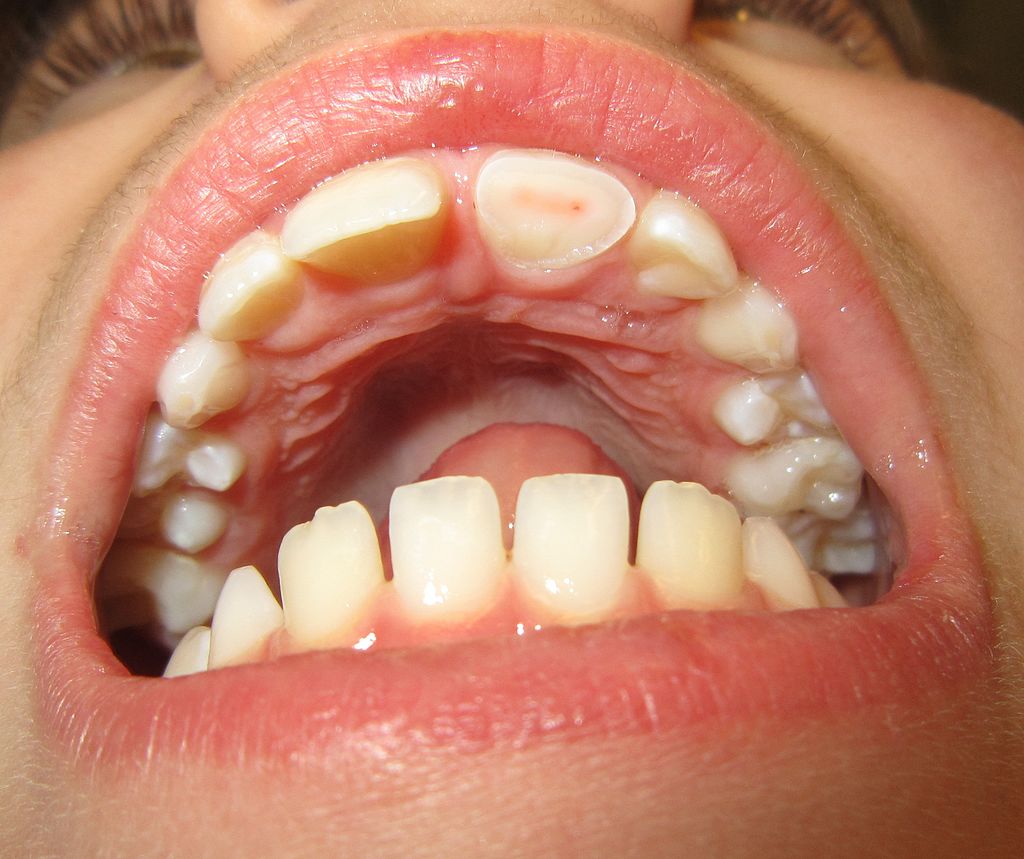
Whether the inciting trauma will result in a tooth fracture, luxation, or avulsion depends on multiple factors, including the location of the tooth in the dental arch, the force and direction of the impact, the structure of the impacting object, and the integrity of the tooth and its surrounding structures.[8] Teeth located anteriorly in the maxilla are most likely injured because of their position within the oral cavity. Any patients with previous dental injury, dental caries, or inflammatory changes of the tooth’s surrounding structures are more predisposed to traumatic injuries with even a low impact than those with healthy tissues.[9]
It takes a significant amount of force to avulse a tooth; thus, additional injuries to the surrounding alveolar bony structures and the entire head and neck should be considered in patients with avulsed teeth.[2]
Infectious Dental Emergencies
Dental caries are the basis for the majority of dental infections. While dental caries themselves are not life-threatening, their presence can predispose patients to infections of the oral cavity. Other causes of dental infections include trauma or operative manipulation such as root canal treatment and third molar extractions.[10]
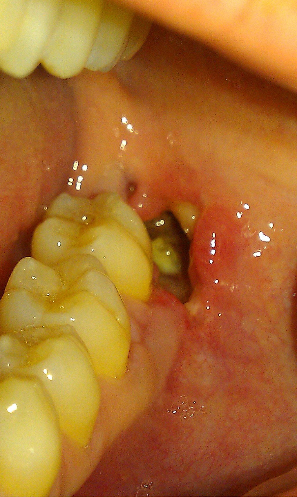
Infections of dental origin prompt patients to seek emergency treatment mainly due to severe pain or swelling. These include irreversible pulpitis, pulp necrosis, periodontitis, and apical and periodontal abscesses. While periodontal disease does not normally induce pain, a severe necrotizing form of the disease exists, which is characterized by an aggressive infection with extreme gingival pain.[11] Pericoronitis, the infection of the gingival tissue overlying an erupting or partially erupted tooth, and typically a 3rd molar may also cause localized pain and swelling.
The progression of dental infections can lead to deep space infections of the head and neck and osteomyelitis of the facial bones. In one study, dental infections were the most common cause of almost 50% of deep neck abscesses.[12] Likewise, dental infections were found to be the etiology for over 90% of patients who presented with Ludwig angina, a rapidly progressive and sometimes fatal infection of the soft tissues of the mouth and neck, which can ultimately cause airway compromise.[13] Thus, early identification and prompt treatment of dental infections are important to prevent more severe, widespread, and life-threatening infections.
Post-procedural Dental Emergencies
Post-procedural Bleeding
Hemostasis may be disrupted by multiple mechanisms, which can be organized into local and systemic etiologies. Hemostasis can be disrupted locally when the blood clot fails to form or is lost at the site of vessel injury. This depends on intrinsic factors, including the surgical location (whether the site is easily accessible) and wound size, with smaller lesions responding better to local treatment. Extrinsic factors, including accidental disruption of the clot through persistent spitting or rinsing, as well as constant suctioning, can also affect local hemostasis.[14]
A systemic disruption of the hemostasis is seen in congenital and acquired coagulopathies. Congenital coagulopathies are rare, but the most commonly encountered are hemophilia A, hemophilia B, and Von Willebrand Disease, which inhibit stable clot formation.[15] Acquired coagulopathies include patients with systemic diseases that affect either the clotting factors or platelet number and integrity. This commonly involves patients with cirrhosis or end-stage renal disease managed with dialysis.[14] Acquired coagulopathies also include patients on anticoagulant medications such as enoxaparin, warfarin, and direct oral anticoagulants, all of which affect the clotting cascade in unique ways and thus prevent stable clot formation.
Alveolar Osteitis
While the etiology of alveolar osteitis is not fully understood, many hypotheses have been studied to determine why clot formation at the extraction site either doesn’t develop or prematurely breaks down. The etiology is likely multifactorial as many studies have found both physiologic and non-physiologic mediators which promote premature clot lysis. Although poorly understood, cytokines (specifically IL-6), biomarkers, local intraoral bacteria, and poor oral hygiene have all been implicated as potential culprits in the pathogenesis of this disease.[7]
Epidemiology
Traumatic Dental Emergencies
Oral trauma accounts for a relatively low percentage of traumatic injuries in all age groups but is responsible for around 17% of total injuries seen in children.[16] Dental trauma is more prevalent in males and is more likely to affect permanent teeth than primary teeth. The peak incidence of dental injuries occurs in children in the 7 to 11 age group.[17] Falls account for the majority of cases of dental trauma, approximately 65%. Sports-related dental trauma is most prevalent in the teenage population, with the incidence of physical violence-related dental trauma increasing in young adults ages 21 to 25.[8]
Infectious Dental Emergencies
The three most prevalent orofacial infections in one British study were periapical abscesses (14 to 25%), followed by pericoronitis (10 to 11%), with periodontal abscesses being the least common (6 to 7%).[18] The incidence of periodontitis is dependent on multiple factors, including ethnicity and geographic location; while periodontitis has many subclassifications, it is thought that the general prevalence does not exceed 1%.[19]
Additionally, some dental infections show a preference for distinct populations. For example, since pericoronitis is most commonly seen with erupting third molars, the condition is prevalent in patients in their twenties.[20] Necrotizing periodontal disease, though rare with a worldwide prevalence of less than 1%, is predominantly seen in immunocompromised and severely malnourished individuals. Furthermore, pulp complications are common amongst those that have cracked teeth or dental fractures that go untreated. Wu S. et al. found in a retrospective cohort study that examined individuals with cracked teeth that 65.5% of patients were later diagnosed with irreversible pulpitis, while 34.5% were later diagnosed with pulp necrosis.[21]
Dental infections make up the majority of hospital admissions from the emergency department for those presenting with dental-related complaints. Most people who visit the emergency department with a dental complaint are discharged. Of the 5% who require admission, an overwhelming majority (85%) have a diagnosis of dental infection with diseases of the pulp and periapical tissue.[22]
Post-procedural Dental Emergencies
Post-procedural Bleeding
Bleeding is a widely recognized and expected complication encountered in dental practice, and the incidence increases with the presence of congenital or acquired coagulopathies.[5][23][24][25] The incidence of post-extraction bleeding ranges from 0% to 26%, increasing the incidence in patients using anticoagulant medications.[26] With anticoagulant use on the rise in the last decade, these patients are more likely to present to the dental setting than patients with congenital coagulopathies.[27] Bleeding rates are higher in patients using warfarin when compared to patients using direct oral anticoagulants.[26]
While the prevalence of patients with congenital coagulopathies remains low, hemophilia A, hemophilia B, and Von Willebrand disease account for 97% of this population, with Von Willebrand disease being the most common.[15][28] While many patients present with a known congenital coagulopathy diagnosis, up to 14% of patients with hemophilia might not be diagnosed until after sustaining a severe bleeding episode with routine dental treatment.[15]
Prolonged postoperative bleeding most commonly occurs in the mandibular molars compared to the maxillary molars. Dental extractions involving infected supporting structures are also associated with an increased incidence of post-extraction bleeding.[5]
Alveolar Osteitis
While the incidence of alveolar osteitis ranges between 0.5% and 68% in the current literature, studies with larger patient populations suggest that the true incidence is likely less than 5%.[6][29][30] However, the incidence does appear to be higher in lower third molar extractions, with up to 30% of these patients developing alveolar osteitis.[31] Other risk factors include poor oral hygiene, tobacco use, oral contraceptive use, and difficult extraction.[7]
Pathophysiology
While pertinent pathophysiology differs for each dental emergency, a basic understanding of the anatomy and structure of the teeth and their surrounding structures sets the foundation for understanding the impact of the disturbances of normal physiology for each category of dental emergencies.
Each tooth consists of a crown, the visible portion, which is connected to the root, or the portion of the tooth that extends into the gingiva and alveolar bone. The enamel is the outermost layer of the tooth and overlays the dentin layer. Within the dentin layer is the pulp, which houses the neurovascular supply that extends from the alveolar bone through the tooth’s root.[10]
The periodontium consists of four main components that comprise the surrounding structures that support dental health: the gingiva, periodontal ligament, alveolar bone, and cementum. The gingiva (i.e., the gums) is the specialized mucosa that surrounds and protects the teeth. The periodontal ligament connects the alveolar bone to the cementum tissue, which covers the tooth root.[32] The normal oral cavity flora consists of many bacteria, especially gram-negative and facultative anaerobes.[10]
Traumatic Dental Emergencies
In tooth luxations and avulsions, the periodontal ligament can partially or fully tear, allowing for a partial or complete displacement of the tooth from its alveolar socket. As the neurovascular supply for each tooth comes from the alveolar bone, disruption of this periodontal ligament can ultimately lead to pulp necrosis, infection, and tooth loss. The fibers of the periodontal ligament are delicate and may not fully recover even with the replantation of an avulsed tooth. Resorption of the root may occur, further compromising the neurovascular supply, and leading to dental fractures and tooth loss.[33][34]
Infectious Dental Emergencies
Pulpitis and Pulp Necrosis
The pulp is the center part of a tooth directly beneath the dentin layer, which contains nerves and blood vessels. Bacterial decay can erode through the enamel and dentin layers and enter the pulp; this causes an infection of this tissue resulting in pulpitis. The microbes causing pulpitis continue to be an area of study, but it is predominantly thought that anaerobic bacteria are the leading cause of pulp infections.[35] Pulp necrosis results when the pulpitis infection progresses and causes irreversible damage and cell death.
Periapical Abscess
The apex of a tooth is the area where the nerve enters the tooth through the root tip. Bacterial infections of the pulp can progress and collect in the periapical region, creating an abscess. Initially, this abscess is small and focal to the involved tooth. However, local extension of this infection can lead to serious complications.
Periodontitis and Necrotizing Periodontal Disease
Periodontitis is an infection of the pocket surrounding the base of the tooth and gum tissue. It can almost universally be attributed to untreated dental caries, which spread to the gingival epithelium. Advanced periodontitis can lead to irreversible loss of tooth attachment and alveolar bone loss.[36]
Periodontal disease is a progression from gingivitis and involves transition and accumulation from aerobic bacteria to anaerobic bacteria such as Aggregatibacter actinomycemcomitans and Porphyromonas gingivalis. This fosters a host inflammatory response that further contributes to the condition. While periodontal disease is not considered a dental emergency, it becomes emergent when it progresses into a necrotizing infection. While the pathogenesis of necrotizing periodontal disease is not entirely understood, it is largely thought that a reduced host immune response allows commensal oral organisms to become pathogenic. These bacteria produce several metabolites that subsequently cause the rapid destruction of the periodontium.[11]
Periodontal Abscess
A periodontal abscess occurs when there is an obstruction of the periodontal pocket, causing the accumulation of bacteria. These abscesses involve the supporting structures of the teeth, including the periodontal ligaments and alveolar bone. Blockage can be caused by foreign body impaction, e.g., toothpick or dental floss, or calculus accumulation. The local inflammatory response to the abscess formation, such as lysosomal enzyme release from host neutrophils, contributes to tissue damage in addition to bacterial accumulation.[37] Not surprisingly, there is a positive correlation between the development of a periodontal abscess with the depth of the periodontal pocket.[38]
Pericoronitis
Pericoronitis is an infection of the gingival tissue surrounding an erupting or partially erupted tooth. Before the eruption, the crown of the tooth exists in a sterile space of the dental follicle. Eruption requires exposure through the gingiva into the oral cavity, which contains a flourishing microbial environment. A small pocket can form between the gingiva and the grooves of the erupting tooth, making it difficult to clean and creating an area where food can easily be impacted. This creates an ideal environment for bacterial overgrowth to progress to an infection of the surrounding soft tissues.
Interestingly, the microbial composition of pericoronitis is thought to be different than that of periodontitis.[39] Although pericoronitis can occur in any tooth, it is most commonly associated with the eruption of wisdom teeth, likely due to the posterior position of this tooth in the mouth and the deep grooves of this particular tooth.[20]
Post-procedural Dental Emergencies
Post-procedural Bleeding
When the vascular endothelium is injured during a routine dental procedure, hemostasis starts by triggering the clotting cascade and forming a platelet plug. While gingival bleeding may occur after dental extractions, the major source of bleeding is the alveolar bone. Local soft tissue bleeding is more likely to occur with a traumatic extraction, which can result in lacerated blood vessels. Osseous bleeding can be from the central vessels or nutrient canals.[5]
Bleeding in hemophilia A and B occurs because of defective fibrin stabilization due to deficiencies in factors VIII and IX, respectively.[40][41] Furthermore, the Von Willebrand factor increases the half-life of factor VII, and thus patients with Von Willebrand disease will also have defective fibrin stabilization due to a similar mechanism as hemophilia A.[42]
Bleeding in patients on anticoagulation therapy occurs because of the inhibition of specific clotting factors, depending on the medication used. Enoxaparin specifically potentiates antithrombin III, which in turn inactivates factor Xa.[43] Warfarin depletes vitamin K-dependent factors and thus reduces the synthesis of factors II, VII, IX, and X.[44] Direct oral anticoagulants are a class of medications that specifically inhibit factor Xa or IIa (thrombin).[45]
Liver and renal pathology can also lead to hematologic consequences. Patients with cirrhosis can experience pancytopenia due to hypersplenism, which can affect platelet plug formation during hemorrhage. Additionally, cirrhosis also leads to impaired coagulation, with the vitamin K-dependent factors being the most affected.[46] Patients with end-stage renal disease also have increased bleeding tendencies because of uremia-induced platelet dysfunction and are more likely to have chronic normocytic anemia at baseline.[47]
Alveolar Osteitis
The pathophysiology is not well understood, but it is generally accepted that either physiologic or non-physiologic mediators trigger premature clot lysis, which ultimately results in alveolar bone exposure after exodontia. Because the literature has shown many factors significantly affect clot formation and lysis, alveolar osteitis is likely a multifactorial phenomenon. Some areas of the previous study include cytokines (specifically IL-6), several biomarkers, local intraoral bacteria, and poor oral hygiene, all of which have been implicated as potential culprits in the pathogenesis of this disease.[7]
History and Physical
Traumatic Dental Emergencies
Tooth Fractures
The classification of dental fractures is dependent upon the tissues involved.[9] Fractures from the most superficial to deepest include enamel infractions, enamel fractures, enamel-dentin fractures, enamel-dentin fractures with pulp exposure, crown-root fractures., and root fractures.
Enamel infractions are the least worrisome and involve microcracks isolated to the enamel. The tooth structure is preserved, and patients are usually asymptomatic. The enamel abnormalities are visualized by transillumination of the teeth during the physical exam. In contrast, enamel fractures involve more substantial damage to the enamel, yet they still do not involve the dentin or pulp layers.[48]
Both classifications have a normal response to pulp vitality tests and tooth mobility testing; however, radiographs of enamel fractures will demonstrate the degree of enamel loss, while enamel infractions will have normal imaging.[49]
Enamel-dentin fractures extend through the enamel and into the dentin without exposing the pulp layer. Enamel-dentin fractures with pulp exposure are more complex in that the fracture extends to the pulp layer, and the pulp is visible on physical exam.[48]
For enamel-dentin fractures, the physical exam will usually demonstrate a vital tooth without sensitivity to percussion and normal mobility testing. In contrast, enamel-dentin fractures that extend to the pulp will clinically be sensitive to applied pressure and temperature testing, yet the pulp is generally viable unless there is a concomitant luxation injury.[49]
Crown-root fractures extend from the tooth’s crown toward its apex and involve the cementoenamel junction overlying the root. The pulp may or may not be involved. The degree of apical involvement is difficult to visualize as the apex of the tooth is below the gingival margin; however, the tooth will likely be mobile, and the patient will also complain of sensitivity to percussion or pressure.[48] Radiographs with multiple views (i.e., a parallel periapical view, additional views with vertical and horizontal angles, and an occlusal view) are recommended as they can better reveal the extent and location of the apical fracture.[49]
Root fractures involve the cementum, dentin, and pulp. Again, the fracture is below the gingival margin and can be difficult to visualize during the physical exam. However, there may be clinical gingival bleeding, percussion pain, and crown mobility.[48] Radiographs with multiple views (i.e., a parallel periapical view, additional views with vertical and horizontal angles, and an occlusal view) are integral to better evaluate the fracture.
Tooth Luxations
In the setting of dental trauma, a physical exam demonstrating a tooth with abnormal loosening is consistent with tooth subluxation. If a tooth is found to be displaced out of its alveolar socket in an incisal/axial direction, it is considered to be an extrusive luxation. This is in contrast to an intrusive luxation which involves the displacement of the tooth apically into the alveolar bone. Lateral luxations consist of a tooth displaced in any lateral direction and are commonly associated with fractures involving the alveolar socket wall.[49]
Tooth subluxations are often tender to light touch and percussion, have increased mobility without displacement and may have minimal gingival bleeding. Because there can be transient pulp damage, pulp sensitivity testing may initially be negative. If obtained, radiographs are generally normal.[49]
With extrusive luxations, the tooth appears elongated incisally or displaced in the direction of the crown. The tooth will have increased mobility and negative pulp sensitivity tests. On radiographs, the tooth will have an increased periodontal ligament space apically and appear displaced incisally compared to the adjacent non-injured teeth.[49]
With intrusive luxations, the tooth will appear to be displaced apically into the alveolar bone. Because the tooth is displaced within the alveolar bone, it will be immobile and likely negatively respond to pulp sensitivity testing. On radiographs, the tooth will have a partially diminished or completely absent periodontal ligament space and appear to be displaced apically compared to the adjacent non-injured teeth.[49]
With lateral luxations, the tooth can be displaced in any lateral direction, further described as buccal, lingual, mesial, and distal directions. Lateral luxations are often associated with an alveolar bony fracture which can entrap the tooth’s root making mobility testing results variable. Radiographs will demonstrate a widened periodontal ligament space which may only be seen if additional horizontal views are obtained.[49]
As it takes a significant amount of force to cause an extrusive, intrusive, or lateral tooth luxation, a thorough examination of the oral cavity should be performed to evaluate for other traumatic injuries, including lacerations, gingival contusions, and mandibular or midface fractures. Radiographs may be useful for assessing the exact positioning of the tooth and will also aid in identifying the extent of the injury.
Tooth Avulsion
In the setting of dental trauma, a physical exam demonstrating a missing tooth from its alveolar socket is consistent with a tooth avulsion. It is essential to obtain history regarding the length of time since the tooth was avulsed, whether the involved dentition is a primary or secondary tooth, whether the patient or family has kept the tooth, and what medium (if any) the tooth has been stored in.
A thorough examination of the oral cavity involves assessing for any other traumatic injuries, including lacerations, gingival contusions, and mandibular or midface fractures. The tooth socket should be evaluated for the presence of tooth fragments (i.e., a sign of dental intrusion) or other foreign bodies that may prevent tooth replantation. If the avulsed tooth is not found, the healthcare provider should consider that the patient may have aspirated or swallowed it and should evaluate for pulmonary versus gastrointestinal foreign body location with radiographs.
Infectious Dental Emergencies
Identifying dental infections is often straightforward and obvious from the history and physical examination—most patients with early dental infections present with complaints of localized pain or swelling.
Patients with reversible pulpitis usually present with a severe toothache exacerbated by temperature changes; as the pulp vitality further deteriorates into irreversible pulpitis and pulp necrosis, dental pain becomes more constant. As pulp necrosis develops, the tooth becomes discolored and can become yellow, gray, brown, or black in appearance.
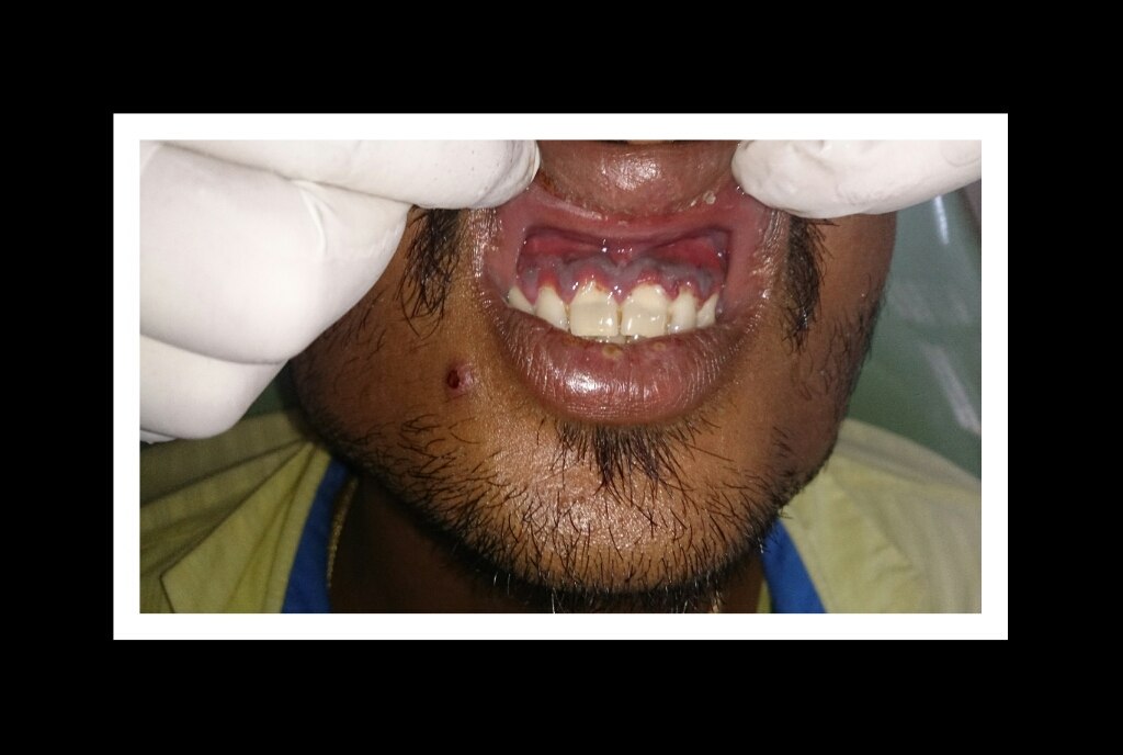
Those with periodontitis experience gingival pain, bleeding after brushing their teeth, as well as halitosis. Furthermore, gingival inflammation is present during the oral examination. Usually, patients are known to have deep periodontal pockets (4 -12 mm) though these are often not able to be measured during an acute infection.[50]
Patients with necrotizing periodontal disease also present with severe pain and halitosis. However, they often report a rapid onset on systems, and the periodontal attachment can be destroyed in just a few days.[11] The physical exam will be notable for “punched-out” ulcerations, necrotic gingiva, and pseudomembrane development consisting of inflammatory cells. Patients often experience spontaneous gingival bleeding or bleeding with minimal manipulation. Unlike periodontitis, deep periodontal pockets are rare, given that the quick progression of the disease does not allow enough time for pocket formation.[11]
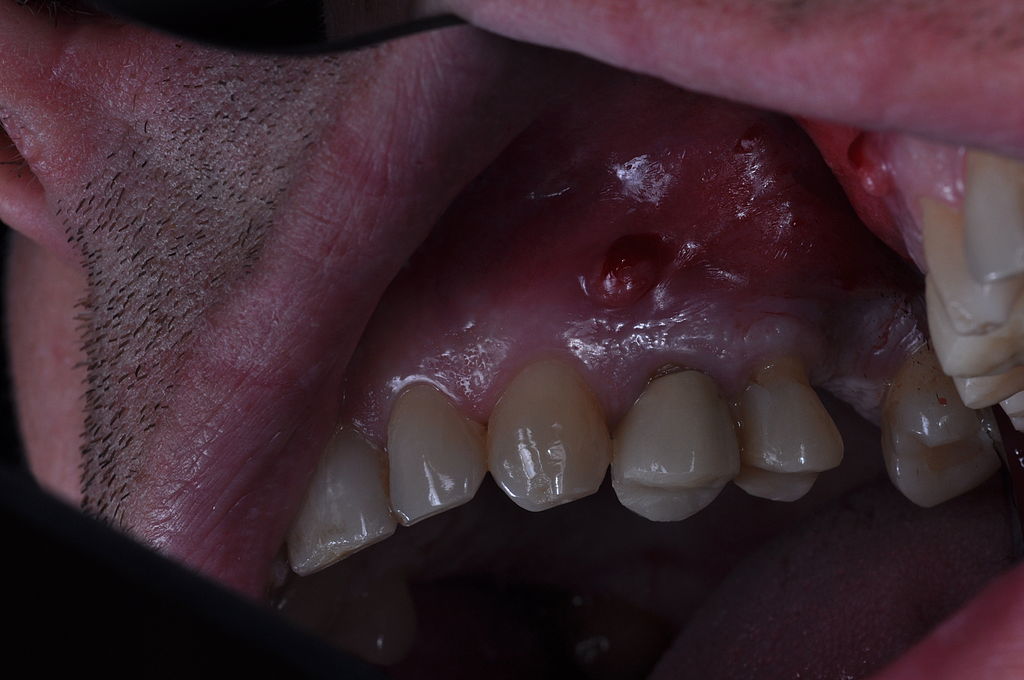
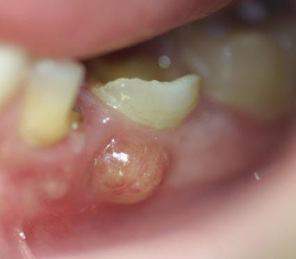
Periapical abscesses usually have a palpable swelling at the base of the tooth.[51] Similarly, periodontal abscesses typically present as an intraoral swelling with or without pain. These patients may complain of pain exacerbated by biting or tooth laxity. This is due to the gradual loss of the periodontal ligament. A purulent discharge may be elucidated by biting or probing.[52]
Pericoronitis is associated most often with third molar eruptions, and patients complain of pain in the posterior mandible or maxilla. Inspection reveals a fully erupted or partially erupted tooth with surrounding gingival tissue that is erythematous and inflamed.[20]
More serious facial and deep neck, mediastinal, or brain infections can arise from untreated dental infections and have more complicated and serious presentations.
Post-procedural Dental Emergencies
Post-procedural Bleeding
Immediately after a tooth extraction, bleeding commonly occurs. Normal postoperative bleeding is most often described as a slow ooze, and clot formation generally occurs within several minutes, and the majority of the bleeding should resolve within 30 minutes. It is common for patients to experience minimal oozing and blood-tinged saliva for up to 8 hours after the extraction.
Post-extraction bleeding can be further classified into primary, reactionary, and secondary prolonged bleeding. Primary prolonged bleeding occurs during and immediately after the extraction procedure. In this scenario, the provider will notice the patient’s mouth actively filling with blood following the removal of a hemostatic dressing. Reactionary prolonged bleeding occurs several hours after the dental extraction.
Patients with reactionary prolonged bleeding are more likely to be on anticoagulation therapy, have a systemic illness, or have a congenital coagulopathy. Secondary prolonged bleeding generally occurs one week after the extraction and is rarely encountered in dental practice. Secondary infections are the likely etiology of prolonged secondary bleeding. Primary prolonged bleeding is more likely to respond to local treatment measures such as pressure packs and hemostatic agents, whereas reactionary prolonged bleeding may require a systemic intervention.[5]
Post-extraction bleeding may also result in local oral hematoma development, which would be apparent on the patient’s physical exam. The hematomas may expand over time resulting in airway compromise, and any hematomas should be monitored frequently for progression.
In the event of a traumatic extraction, the provider should perform a thorough physical exam to evaluate for any lacerations or other soft tissue damage that would require repair.[5]
Alveolar Osteitis
Patients with alveolar osteitis generally present one to five days after a dental extraction with severe pain at the extraction socket. This pain can radiate in the trigeminal nerve distribution and is unrelieved by oral analgesics.[7]
The physical exam will demonstrate expected postoperative changes with a partial or complete loss of the blood clot within the alveolar socket. Though it can be difficult to appreciate on visual inspection, exposed alveolar bone is the classic physical exam finding of alveolar osteitis.[53]
Evaluation
Traumatic Dental Emergencies
Both intraoral and extraoral radiographs, in addition to CT scans, can be used to help identify dental and periodontal fractures. The recommended radiographs are one parallel periapical view, two additional views with a vertical and horizontal angle, and an occlusal view.[49]
They can be useful to discern between a dental luxation and a dental avulsion and can also better evaluate the extent of fractures below the gingival margin. Depending on the history obtained and the physical exam, radiographs and CT scans may not change the treatment plan. Dentists should consider the risks and benefits of radiation exposure to the patient.[17][9]
Infectious Dental Emergencies
Visual inspection and palpation are the best initial methods to evaluate for infection. Common signs of infection include tissue erythema, swelling, or pus extrusion. Tooth discoloration and gingival necrosis may also be noted. Periodontal probing can aid in the diagnosis of periodontal disease.
Dental radiographs can show the extent of the infection and can help evaluate areas not obvious to visual evaluation. These include intraoral periapical radiographs, bitewing radiographs, and panoramic radiographs. These radiographs help to identify the extent of dental caries, the presence of a periapical abscess, alveolar bone loss concerning periodontitis, as well as third molar eruptions. All radiographic evidence must be confirmed with the dental practitioner’s physical examination for a full evaluation and accurate diagnosis.
CT scans and MRIs help assess deep-space infections and osteomyelitis. Cone-beam CT is becoming increasingly common in clinical settings to visualize areas of oral infections better.[54]
Post-procedural Dental Emergencies
Post-procedural Bleeding
Unless in a traumatic oral injury setting, radiographs and CT scans have little utility in managing post-extraction and postoperative bleeding.
Ideally, any history of congenital coagulopathy, systemic illness, or medication use that would predispose a patient to have abnormal bleeding would have been identified by the oral surgeon or dentist before the procedure. It can be useful to ask whether bleeding disorders are present in any members of the patient’s family, as congenital coagulopathies are inherited and may not yet be diagnosed in the patient themselves. If a patient has been diagnosed with congenital coagulopathy, it is ideal for coordinating their dental care in conjunction with their hematologist and providing factor replacement before any invasive procedures to prevent postoperative bleeding. Similarly, if a patient is known to have a systemic illness or uses anticoagulant medications, it can be beneficial to coordinate their dental care in conjunction with their primary care physicians to minimize the risk of postoperative bleeding.[28]
Some literature reviews suggest that patients should discontinue their anticoagulation medications before their dental procedure. Clinical trials suggest that patients on warfarin or direct oral anticoagulants would benefit from starting oral tranexamic acid before their dental procedure. However, these points remain controversial, and other sources suggest that the risk of bleeding is lower than the risk of thromboembolism and recommend patients continue their anticoagulant therapy perioperatively given the high efficacy and availability of local hemostatic therapies.[23][25][55][56][57]
In addition to a thorough physical exam, laboratory studies can be performed preoperative or postoperative if clinically indicated to evaluate hemoglobin and platelet levels, to identify supratherapeutic international normalized ratio (INR) levels with warfarin use and in patients with cirrhosis, and to determine clotting Factor VIII and IX blood levels in patients with Hemophilia A and B, respectively.
Alveolar Osteitis
Alveolar osteitis is a clinical diagnosis and does not require laboratory studies or radiographic examination. A thorough history and physical exam are sufficient to diagnose this postoperative complication. However, if there is a concern for a retained tooth fragment within the alveolar socket, panoramic radiographs can be performed.[7]
Treatment / Management
In addition to the treatments described below, all dental emergencies benefit from basic oral hygiene measures to prevent the overgrowth of the natural oral cavity flora and prevent opportunistic infections.
Traumatic Dental Emergencies
Tooth Fractures
Enamel infractions generally do not require treatment. However, if the microcrack starts progressing in size, etching and sealing with bonding resin is recommended to prevent discoloration and bacterial infection.[49]
If the tooth fragment is available, enamel fractures can be managed by reattaching the enamel fragment. If the tooth fragment is unavailable, the tooth edges can either be smoothed if the lesion is small or restored with resin composite if the lesion is more extensive. It is recommended that patients follow up in 2 and 12 months so the dentist can assess the integrity of the restoration clinically and with radiographs.[49]
The focus of initial treatment for enamel-dentin fractures is to protect the exposed dentin. Calcium hydroxide liners are commonly available in urgent care clinics, emergency departments, and primary care offices. These healthcare providers should apply this temporizing treatment to prevent infection and protect the tooth’s viability.[58] In dental offices, calcium hydroxide can also be applied initially; however, the material eventually needs to be covered with a composite resin or glass ionomer. If the fragment is available, a dentist can consider reattaching it. If the fragment is unavailable, the dentist can perform restorative procedures using various materials, including resin direct composites or ceramics.[49]
The focus of initial treatment for enamel-dentin fractures with pulp exposure is to protect the pulp. Calcium hydroxide is one method to accomplish this and is often available in non-dentistry practice settings, and when initiated early, it can help prevent bacterial infection.[58] For definitive treatment, the dentist has two main avenues of managing pulp exposure: pulp capping or partial pulpotomy. The best approach is dependent on the degree and duration of the pulp exposure and the presence of other injuries.
Pulp capping works best in immature teeth with open apices in which the pulp was considered to be healthy before the trauma. Indications for pulp capping include pulp exposure of a small diameter (less than 1.5 mm) that has only been exposed for a short amount of time, thus minimizing the risk of bacterial invasion.[48]
Partial pulpotomy is indicated when either the duration of pulp exposure has been extensive, or the diameter of exposure exceeds 1.5 mm. In these cases, a partial pulpotomy can be performed, which ultimately allows the tooth to remain vital for an extended amount of time. However, the tooth will eventually require root canal therapy and a subsequent restoration procedure.[9]
Crown-root fractures are initially managed by identifying the extent of the fracture and exposing its margins. If still attached, the tooth fragment is removed by the dentist to evaluate for pulp involvement. If the pulp is not involved, the fragment can be reattached, or the exposed dentin can be covered with resin composite or glass ionomer.
If the pulp is exposed or the fracture extends far past the cementoenamel junction, definitive management becomes more complex and interdisciplinary, involving the dentist, endodontist, and oral surgeon.[9][49]
The initial focus for treating root fractures involves ensuring appropriate crown fragment placement on physical exams and radiography. The crown will often be mobile and thus will need to be stabilized with a dental splint for weeks to months, with cervically located fractures requiring a longer duration of immobilization when compared to apically located fractures.[49]
It is recommended that patients who have undergone treatment for enamel-dentin fractures with pulp exposure, crown-root fractures, and root fractures should have regular follow up with their dentist or oral surgeon at two months, three months, six months, and 12 months duration.[49]
Tooth Luxations
Tooth subluxations generally do not require treatment; however, a passive and flexible splint can be utilized to stabilize the tooth if there is marked mobility or tenderness when the patient bites down. If a splint is applied, it is recommended that the patient follow up with a dentist after two weeks to remove the splint. If a splint is not utilized, the patient should be reevaluated by a dentist clinically and radiographically after two weeks, 12 weeks, six months, and 12 months.[49]
Extrusive luxations require that the tooth be gently pushed back into its alveolar socket under local anesthesia. The tooth needs to be stabilized with a passive and flexible splint for two to four weeks. The patient is recommended to follow up with a dentist after two weeks, four weeks, eight weeks, 12 weeks, six months, and 12 months. The patient will require endodontic treatment if subsequent radiographs or pulp sensitivity testing indicate pulp necrosis.[49]
Intrusive luxations in teeth with incomplete root formation may spontaneously re-erupt without intervention. If spontaneous repositioning does not occur within four weeks, orthodontic repositioning becomes necessary. Teeth with complete root formation may spontaneously re-erupt if there is less than 3 mm of intrusion; however surgical repositioning should be initiated if there is no spontaneous eruption within eight weeks. If there is more than 3 mm of intrusion, the tooth should be repositioned surgically. For four weeks, the tooth should be stabilized in a passive and flexible splint. While teeth with incomplete root formation are unlikely to develop pulp necrosis, mature teeth almost always experience pulp necrosis, and the health of the pulp should be monitored at subsequent follow-up appointments. Root canal therapy should be considered early.[49]
Lateral luxations require that the tooth be gently relocated back to this alveolar socket under local anesthesia. This can be difficult in certain situations when the root is “locked” into position by a concomitant alveolar fracture and may require the dentist to first disengage the tooth apex before repositioning it. The tooth will require stabilization with a passive and flexible splint for at least four weeks, perhaps longer if there are concomitant alveolar fractures. Two weeks after the injury, the pulp should be evaluated for pulp necrosis with sensitivity testing. Teeth with incomplete root formation may experience spontaneous revascularization. However, teeth with complete root formation will likely experience pulp necrosis, and root canal therapy is often indicated. The patient is recommended to follow up with a dentist after two weeks, four weeks, eight weeks, 12 weeks, six months, and 12 months.[49]
Tooth Avulsions
If possible, replantation is the preferred treatment modality.[59] This must occur quickly, with the highest tooth survival seen with tooth replantation within the first 30 minutes. Early treatment focuses on preserving both tooth viability and a functioning alveolar socket.[17] Primary teeth are never replanted, as this can affect the normal eruption of underlying permanent teeth.
Before replantation, it is recommended that the avulsed tooth be stored in an isotonic solution to prevent apoptosis of the periodontal ligament. Such acceptable solutions include milk, normal saline, or the patient’s saliva. Cell death of the periodontal ligament is inevitable and can only be temporarily delayed with a proper storage medium.[60] The alveolar bone should be gently irrigated with normal saline, and any obstructing blood clot should be aspirated before replantation.
The tooth should be replanted in the correct anatomic alignment with firm pressure into the alveolar socket. It should then be stabilized with a passive and flexible splint.[9] Immobilization with a splint allows the periodontal ligament fibers to reform connections between the alveolus and the cementum. It is recommended that patients follow up to have their splint rechecked and/or removed at two weeks. If, at this time, the tooth is not viable, root canal therapy is indicated. If the tooth is viable, radiographs should be obtained at the 2-week, 1-month, 3-month, and 6-month follow-up appointments to evaluate for resorption of the tooth. Root canal therapy or even tooth extraction may be indicated if resorption develops.[49]
Infectious Dental Emergencies
Dental infections should be promptly identified and treated as soon as possible to avoid contiguous or hematogenous spread and subsequent life-threatening complications.
As with any infection, complete removal of the source is always ideal but not always practical in the case of dental infections. Tooth extraction is an option for certain patients but is usually the last resort. Often other techniques are recommended so patients can maintain their dentition.
Pulpitis requires treatment aimed at pulp protection and insertion of restorative materials if considered reversible. Irreversible pulpitis and pulp necrosis are treated by either root canal therapy or tooth extraction.
Periapical abscesses and periodontal abscesses require incision and drainage if a tooth extraction is not the desired option. Oral antibiotics should also be given if there is surrounding cellulitis or the patient is immunocompromised. Antibiotics should include gram-negative bacteria and anaerobic coverage. Common antibiotic choices include amoxicillin, amoxicillin-clavulanate, or clindamycin.[61] Large abscesses and those that involve deep space infections require hospital admission, parenteral antibiotics, and sometimes operative intervention.
Treatment for periodontitis is almost universally non-surgical. Mitigation of risk factors such as tobacco cessation and diabetes control is essential for treatment. Professional dental cleanings can be a first step to reversing the disease as it removes the gross buildup of plaque and calculus. Proper home oral hygiene and antimicrobial mouth rinses such as chlorhexidine gluconate are recommended. Rarely systemic oral antibiotics are needed. In extreme cases, surgery to remove the infected gums can be done to maintain dentition.[36]
Treatment for necrotizing periodontal disease is dependent on the severity of the disease. However, the initial management generally involves mechanical debridement of necrotic tissue under local anesthesia, removal of any pseudomembrane, and aggressive oral hygiene incorporating an antiseptic mouthwash. In addition to these local measures, oral antibiotics are indicated if systemic symptoms (such as fever or lymphadenopathy) are present. Mitigating risk factors such as tobacco use and/or any underlying immunologic condition is also important in managing necrotizing periodontal disease.[11]
Treatment for pericoronitis includes non-surgical and surgical treatment options. Aggressive oral hygiene with irrigation of the pericoronal space is recommended to dislodge any impacted debris.[62] Often oral antibiotics are helpful in decreasing the bacterial load in that area.[63] Surgical treatment involves removing the soft tissue layer overlying the erupting third molar, which is called the operculum. This procedure is beneficial as it not only removes the deep pocket within the gingiva but also makes it easier for the tooth to erupt, as well.[64] Extraction of the involved tooth is often an acceptable treatment for pericoronitis involving third molar eruptions.
Post-procedural Dental Emergencies
Post-procedural Bleeding
The initial treatment of post-extraction and postoperative bleeding is to augment hemostasis while maintaining airway patency. The interventions available for post-extraction and postoperative bleeding can be categorized into local or systemic treatments.[5]
Local interventions consist of both surgical and non-surgical hemostatic measures. Basic non-surgical hemostatic measures include manual pressure or pressure packing with gauze. However, an array of therapies can be applied locally to either absorb or halt bleeding. These therapies typically involve hemostatic pharmacologic or biologic agents that are impregnated into packing material, sponge, gel foam, adhesive, or solution, which are applied locally to the site of bleeding. These therapies have been well documented in the literature and are considered effective in common practice.[5][55] Below is a list of common locally acting hemostatic agents organized by category:
- Adhesive agents: cyanoacrylate glue, fibrin glue, and chlorhexidine bio-adhesive gel
- Absorbable agents: oxidized cellulose, gel foam, collagen fleeces or sponges, and gelatin sponge
- Biologic agents: topical thrombin, green tea extract, Haemocoagulase, and Ankaferd Blood Stopper
- Sealant agents: acrylic or surgical splints and calcium alginate
- Pharmacotherapies: local antifibrinolytic solutions (i.e., tranexamic acid mouthwash) and gauze soaked in tranexamic acid
Surgical measures can also be used in isolation or in combination with non-surgical methods to obtain hemostasis. This involves the oral surgeon cauterizing or suturing the site of bleeding. The exact surgical approach will depend on the source of the bleeding.[5]
Systemic interventions should be considered when a patient has a known congenital coagulopathy, a systemic illness that makes them prone to bleeding or using anticoagulant therapies. The role of local hemostatic agents can be limited in these scenarios as they do not address the underlying etiology of the bleeding. Systemic interventions include the administration of:
- Blood products such as fresh frozen plasma (FFP), platelets, or packed red blood cells (pRBCs) in the setting of warfarin use or cirrhosis
- Specific factor replacement therapy in the setting of congenital coagulopathies
- Anti-hemophilic factor A or anti-hemophilic factor B for Hemophilia A and B, respectively
- Von Willebrand factor concentrate on the setting of Von Willebrand disease
- Prothrombin complex concentrate (PCC) in the setting of cirrhosis, warfarin use, or direct oral anticoagulant use
- Epsilon amino-d-caproic acid in the setting of enoxaparin use
- Desmopressin in the setting of damaged platelets
- Tranexamic acid
Systemic agents should be used cautiously in the anticoagulated patient as there is a risk of thrombotic events with anticoagulation reversal. The risks and benefits of anticoagulation reversal must be considered on an individual basis, discussed with the patient, and using the clinician’s best judgment.[5][25]
The general approach to the patient experiencing postoperative bleeding should be for the dentist or oral surgeon to first clearly visualize the source of bleeding by removing all saliva and residual clot from the surgical site with suction. Manual pressure should be applied firmly with a damp gauze for at least 5 minutes. If the bleeding has not resolved with this measure, local anesthesia should be administered so the patient can be treated with more aggressive therapy. The oral surgeon or dentist should then utilize an appropriate local non-surgical hemostatic agent with or without sutures. If the bleeding is not controlled with these methods, systemic treatment should be considered in conjunction with a hematologist.[5]
If the bleeding is brisk and hemostasis is not obtained such that the patient cannot adequately tolerate their secretions, or if there is an expanding hematoma, the healthcare provider should have a low threshold to secure the patient’s airway in conjunction with definitive hemostasis.
Alveolar Osteitis
Not surprisingly, as the pathophysiology of alveolar osteitis is not well understood, there is no consensus on protocols for its treatment or management. Despite this, treatment approaches tend to focus on symptomatic relief rather than addressing any underlying disease process.
While it does little to relieve pain, initial management involves intra-alveolar irrigation, which assists in removing clot debris and necrotic tissue. Local analgesia can be obtained with topical anesthetics gels, which can be applied after irrigation. Local analgesia can also be augmented with oral non-steroidal anti-inflammatory drugs (NSAIDs). Although they may impair healing and cause local inflammatory reactions, there does appear to be a role for the temporary application of medicated dressings (impregnated with analgesic and/or antibacterial compounds) post-operatively. Additionally, chlorhexidine oral rinses after exodontia have been shown to help prevent alveolar osteitis, while its use as a primary treatment is still being investigated.[7]
More recently, low-level laser therapy (LLLT) has been suggested to be an effective alternative treatment with comparable results to other known treatments with the benefit of being both safe and inexpensive.[7] With some studies showing this newer technology to be both effective and advantageous, with further study, it could become a protocolized consensus treatment in the future.
Differential Diagnosis
Traumatic Dental Emergencies
Generally, tooth fractures are accurately diagnosed with a detailed clinical history, physical exam, and radiographic evaluation. However, marked tenderness with manipulation of the tooth or the surrounding structures may indicate the presence of a simultaneous tooth luxation or root fracture.[9]
The differential diagnosis of tooth luxations includes all categories of luxations (i.e., complete tooth intrusion, subluxation, and lateral luxation) in addition to tooth avulsion. Similarly, all tooth luxations should be considered in the differential diagnosis of a tooth avulsion.[17]
Infectious Dental Emergencies
Oncologic etiology can sometimes mimic the appearance of dental infections. Squamous cell carcinoma can contribute to bone loss and be misdiagnosed as periodontal disease.[65] Gingival bleeding can occur from leukemia and also be misdiagnosed as an infectious process.[66] Cysts, tumors, and granulomas can likewise have an infectious appearance.[67] Oral side effects from drug therapy can also be misdiagnosed as an oral infection. Other differentials for dental infection include salivary gland pathology as well as sinusitis.[51]
The limited extent of a localized dental infection must be ensured so as not to miss a more life-threatening related complication such as a deep space neck infection or facial abscess.
Post-procedural Dental Emergencies
Post-operative Bleeding
Traumatic extractions with or without oral lacerations affecting vessels should be considered in the differential diagnosis of post-extraction bleeding and post-operative bleeding. Congenital coagulopathies should be considered in the profusely bleeding dental patient as they may not have been previously diagnosed. Systemic illnesses, such as cirrhosis or end-stage renal disease, or the use of anticoagulants should be considered in bleeding patients when little is known about their medical history.
Alveolar Osteitis
While the diagnosis of alveolar osteitis is straightforward in the setting of an appropriate history and physical exam, if the pain is severe or occurring for longer than one week post exodontia, a broader differential diagnosis should be considered.[29]
In these situations, infectious etiology should be considered, especially if there are signs of tissue abnormalities extending beyond the extraction soc et. Differential diagnoses that should be considered are mandibular or maxilla osteomyelitis, subperiosteal infection, and boney sequestrum.[7]
Prognosis
Traumatic Dental Emergencies
Tooth Fractures
The outcome of a given dental fracture depends on the fracture category, the quality of the treatment received, and whether there was a delay in treatment. The prognosis is favorable for minor fractures involving only the enamel. The prognosis worsens for fractures left untreated or involving deeper structures of the tooth and surrounding tissues; these dental fractures also have a higher likelihood of developing infections.[68]
Tooth Luxations
Tooth subluxations generally have a good prognosis with patients with minimal symptoms, a positive response to pulp sensitivity testing, and immature teeth having the best outcomes.
Extrusive luxations, intrusive luxations, and lateral luxations are prone to worse outcomes in comparison to tooth subluxations. The best outcomes are seen in patients with minimal symptoms, a positive response to pulp sensitivity testing, no marginal bone loss, signs of healing periodontium on radiographs, and skeletally immature teeth. Very symptomatic cases that develop pulp necrosis and experience breakdown of marginal bone or bony resorption have a worse prognosis and often require root canal therapy or endodontic evaluation.[49]
Tooth Avulsions
The prognosis for replanted tooth avulsions depends on several items, particularly the extra-alveolar period. While replantation should be attempted for all avulsed teeth, the outcomes are best for those cases in which replantation occurs within 30 minutes, the tooth was stored in an appropriate storage medium, and there are minimal concomitant dentoalveolar injuries. The success of replanted teeth markedly decreases once the tooth exceeds 60 minutes of extra-alveolar time.[69]
Infectious Dental Emergencies
While dental infections historically carried a high rate of mortality over a century ago, when recognized and treated early, uncomplicated dental infections carry a good prognosis. However, if left untreated, dental infections can spread both contiguously and hematogenously, with the potential to cause numerous complications. The sequela of resultant severe sepsis, septic shock, bacteremia, osteomyelitis, deep neck space infections, mediastinitis, endocarditis, and/or meningitis carries significant morbidity and mortality.[51]
Additionally, studies have shown that teeth and periodontal tissue that experience repeated infections or have preexisting periodontal disease carry a worse prognosis.[70][71] The prognosis is generally improved in situations where risk factors and comorbidities (such as tobacco use, uncontrolled diabetes, and immunological diseases) are addressed and managed appropriately.
Post-procedural Dental Emergencies
Post-operative Bleeding
In most cases, patients experiencing post-extraction and post-operative bleeding do well with local interventions. Patients with congenital coagulopathies tend to have a fair prognosis with local interventions when they are pre-operatively treated with factor replacement therapies. However, some individuals in this population will still experience secondary bleeding and require systemic treatment despite early clotting factor replacement.[28]
While discontinuing anticoagulant therapy remains controversial, several studies suggest that it is safest for patients to continue their anticoagulation therapy for procedures with a moderate risk of bleeding, given the efficacy and availability of hemostatic agents.[23][25][55][56][57]
Alveolar Osteitis
Alveolar osteitis is self-limiting, is not known to cause long-term morbidity, and typically resolves within a few days after initiating treatment. The prognosis without treatment is also excellent as the symptoms still resolve in the absence of treatment in a relatively short duration of time.
Complications
The relevant complications for the various dental emergencies differ depending on the specific diagnosis and category being considered.
Traumatic Dental Emergencies
Complications of traumatic dental emergencies include pulp necrosis, pulpal obliteration, tooth discoloration, dental or periodontal abscesses, fistula formation, and root resorption.[72] Tooth avulsions are especially high risk for loss of pulp vitality, especially as the risk increases with each minute the avulsed tooth remains outside the alveolar socket.[17]
Infectious Dental Emergencies
The most common complication of dental infections is tooth loss. However, if left untreated, dental infections can spread both contiguously and hematogenously, with the potential to cause numerous complications. Through contiguous spread, dental and periodontal infections can lead to osteomyelitis of the mandible or maxilla, deep neck space infections (which include Ludwig angina, retropharyngeal abscess, and mediastinitis), and rarely intracranial infections (which include cavernous sinus thrombosis, meningitis, and subdural empyema). If patients with dental infections become bacteremic, there is the potential for hematogenous spread, which can lead to endocarditis.[51]
Specifically, Ludwig’s angina and retropharyngeal abscesses can compromise a patient’s airway. Patients experiencing Ludwig’s angina may present with soft tissue edema involving the face, oral cavity, and neck, which leads to trismus, respiratory distress, voice changes, and the inability to tolerate oral secretions. In these cases, stridor is an ominous sign, and the provider should have a low threshold to intubate before respiratory distress develops utilizing advanced airway equipment, such as video laryngoscopy and/or fiber optics.[13]
Patients presenting with retropharyngeal abscesses are also ill-appearing and present similarly to patients with Ludwig’s angina; however, they are less likely to have facial or neck edema and are more likely to experience neck stiffness with their discomfort usually exacerbated with cervical extension.[73]
All these sequelae portend high morbidity and mortality, and because their presentations can be varied, healthcare providers must include them in the differential diagnosis for ill-appearing patients experiencing dental infections.
Post-procedural Dental Emergencies
The most frequent complication for patients with post-extraction or postoperative bleeding experience is a return visit to their dental practitioner or the emergency department. They may also develop large intraoral hematomas that rarely can compromise their airway. In cases where hemostasis is difficult to achieve, patients may require blood transfusions or hospitalization.[5]
Alveolar osteitis is self-limiting, and there is no evidence correlating it as a risk factor for developing additional dental complications.
Consultations
If a patient with a dental emergency presents to a practice setting other than an oral surgery clinic or dentistry office (i.e., primary care physician, urgent care clinic, or emergency department), they must be referred to an oral and maxillofacial surgeon or dentist expeditiously. In ill-appearing patients who are unlikely to do well with outpatient treatment, admission to the hospital with an oral maxillofacial surgeon consultation may be warranted.
Traumatic Dental Emergencies
Most dental fractures are safe to treat in the outpatient setting with regular dentistry follow-up. After dental replantation and splinting, tooth luxations and avulsions can also be followed up in the outpatient setting. However, it is not uncommon for patients with these dental emergencies to be admitted to the hospital for other non-dental traumatic injuries.
Infectious Dental Emergencies
Complicated dental infections require either an oral and maxillofacial surgeon or endodontist consultation for expedited definitive treatment. Patients experiencing severe disease, infectious complications, or sepsis may require admission to the hospital for both medical and surgical management of their dental infection(s).
Post-procedural Dental Emergencies
For patients that have a suspected or known diagnosis of congenital coagulopathy, dental care should be managed in conjunction with a hematologist. In patients with systemic diseases that affect hemostasis, such as cirrhosis or end-stage renal disease, dental care should be managed in conjunction with their primary care physician.
Patients experiencing alveolar osteitis should be referred back to their dentist or oral surgeon for definitive management. Further consultation is not required.
Deterrence and Patient Education
Regarding dental emergencies, patient education is specific to the diagnosis and category being discussed.
Traumatic Dental Emergencies
While dental trauma may be difficult to foresee in many circumstances, such as accidental falls, motor vehicle accidents, and physical violence, however, using custom mouthguards in contact sports has been shown to decrease traumatic dental injuries in this specific setting. Patients, parents, school teachers, sports coaches, and first responders should be educated on basic first aid for traumatized teeth. Seeking early medical care is also crucial for successful treatment.[9][17]
Infectious Dental Emergencies
Patients should be educated about the serious sequelae of dental infections and the effectiveness of preventative measures. It is important to counsel patients regarding proper oral hygiene with daily toothbrushing and flossing in addition to scheduling regular dentist visits. This will help prevent dental infections and help dental practitioners recognize and treat dental infections early before complications develop. Furthermore, patients need to be educated on the importance of risk mitigation, including smoking cessation and diabetes control, as these factors can make patients more prone to infectious dental emergencies.
Post-procedural Dental Emergencies
In many cases, post-extraction or postoperative bleeding can be anticipated, and patients should be educated on applying pressure to assist in augmenting hemostasis. Patients with known congenital coagulopathies, a systemic illness that increases bleeding, or anticoagulant use should be informed of their increased risk of bleeding, even with minor dental procedures. Patients with congenital coagulopathy will benefit from having their dental care being managed in conjunction with a hematologist. Dental care should be provided in conjunction with a primary care physician for those patients with cirrhosis, end-stage renal failure, or who are on anticoagulant therapy.
Patients undergoing exodontia should be educated regarding the possibility of alveolar osteitis occurring, especially if they possess a known risk factor associated with an increased incidence, such as tobacco or oral contraceptive use. If the patient is considered to be high risk, prophylactic measures with chlorhexidine treatments can be taken. Patients should be given strict return precautions if they experience symptoms consistent with alveolar osteitis, and dental professionals should assist with expedited follow-up in these situations.
Pearls and Other Issues
Summary of Traumatic Dental Emergencies
| Disease |
Severity |
Evaluation |
Treatment |
| Tooth Fracture |
Emergent |
- Visual inspection and mobility testing to determine the depth of the fracture.
- Radiographs may assist in categorizing the extent of the fracture into the tooth root and/or alveolar bone.
- Pulp vitality to evaluate the viability of the exposed pulp.
|
- Initial treatment consists of applying calcium hydroxide liners, but ultimately this will need to be covered with a composite resin or glass ionomer. Restorative procedures may be required if the tooth fragment is unavailable.
- Pulp capping or partial pulpotomy may be indicated if the pulp is exposed.
- An interdisciplinary approach is recommended for fractures that extend past the cementoenamel junction.
|
| Tooth Subluxation and Luxation |
Emergent |
- Subluxation: tenderness with light touch and percussion with abnormal loosening on physical exam.
- Luxation: displacement of the tooth out of its alveolar socket in any direction (incisal/axial, apically, or laterally).
- Radiographs may assist in evaluating the exact location of the tooth and identifying the extent of the injury.
|
- Initial treatment involves relocating the tooth into its alveolar socket.
- The tooth needs to be stabilized in a passive and flexible splint for 2-4 weeks with regular outpatient dentistry follow-up.
|
| Tooth Avulsion |
Emergent |
- Visual inspection demonstrates a missing tooth from its alveolar socket.
- Radiographs can evaluate for dental fragments or foreign bodies within the alveolar socket.
|
- Replantation is the preferred treatment and is best if done within 30 minutes.
- The replanted tooth should be stabilized in a passive and flexible splint for two weeks.
- Regular outpatient dentistry follow-up is recommended.
|
Summary of Infectious Dental Emergencies
| Disease |
Severity |
Evaluation |
Treatment |
| Dental Caries |
Non-emergent unless extensive disease |
- Physical exam shows demineralization of the enamel subsurface.
- Active carious lesions tend to be white, matte, with rough texture, and are cavitated.
- Inactive carious lesions are brown, shiny, smooth, and nonactivated.
- Radiographs may also assist in detecting caries.
|
- Non-invasive procedures can be attempted for early lesions that can be reached by cleaning implements and that have not invaded far into the dentin.
- Restoration with resin compounds is required for noncleansable cavitated lesions that have invaded the dentin layer.
|
| Pulpitis |
Urgent |
- History of severe pain with temperature changes.
- If reversible, pain is intermittent. If irreversible, the pain is constant.
- Dental caries are evident on exam.
|
- If reversible, initial treatment consists of the insertion of resin materials.
- If irreversible, treatment consists of root canal therapy or tooth extraction.
|
| Pulp Necrosis |
Urgent |
- History of severe pain that is constant.
- The tooth is discolored and can progress from yellow to gray, brown, and then black in color.
|
- Treatment requires a root canal or dental extraction.
|
| Periodontitis |
Urgent |
- History of gingival pain, bleeding after teeth brushing, and halitosis.
- A physical exam will show gingival inflammation with deep periodontal pockets.
|
- Mitigation of risk factors such as tobacco use and uncontrolled diabetes.
- Proper home oral hygiene in connection with professional dental cleanings.
- Rarely requires oral antibiotics.
|
| Periapical Abscess |
Emergent |
- A physical exam will demonstrate a palpable swelling at the base of the tooth without or without evidence of purulent drainage.
|
- Conservative management with incision and drainage is preferred, but dental extraction may be required.
- Oral antibiotics are indicated if there is surrounding cellulitis.
- Inpatient treatment with intravenous antibiotics is indicated if the abscesses are large or have progressed to deep-space infections.
|
| Periodontal Abscess |
Emergent |
- History of pain exacerbated with biting. Purulent discharge may be elucidated by biting.
- A physical exam will demonstrate perioral swelling.
|
- Conservative management with incision and drainage is preferred, but dental extraction may be required.
- Oral antibiotics are indicated if there is surrounding cellulitis.
- Inpatient treatment with intravenous antibiotics is indicated if the abscesses are large or have progressed to deep-space infections.
|
| Necrotizing Periodontal Disease |
Emergent |
- History of rapid development of gingival pain, bleeding, and ulcerations.
- A physical exam will demonstrate "punched-out" gingival ulcerations, gingival necrosis, and pseudomembrane development.
|
- Initial treatment consists of local oral care involving the debridement of necrotic tissue, removal of pseudomembrane, aggressive oral hygiene, and antiseptic mouth rinses.
- Oral antibiotics are recommended if fever, lymphadenopathy, or other systemic symptoms are present.
|
| Pericoronitis |
Urgent |
- History of pain in the posterior mandible or maxilla.
- Physical exam with fully or partially erupted tooth with surrounding inflamed and erythematous gingiva.
|
- Aggressive oral hygiene with irrigation of the pericoronal space.
- Oral antibiotics to decrease the bacterial load.
- Surgically removing the operculum overlying the erupting third molar if conservative measures fail.
|
Summary of Post-procedural Dental Emergencies
| Disease |
Severity |
Evaluation |
Treatment |
| Post-extraction and post-procedural bleeding |
Emergent |
- Bleeding that occurs immediately after and up to several hours after a dental extraction or procedure despite initial hemostatic measures.
- Rarely, bleeding can occur up to one week after the procedure.
|
- Local interventions are utilized first and involve manual pressure, hemostatic pharmacologic or biologic agents, or cautery/suture.
- Systemic inventions may be required if there is a known congenital coagulopathy, a systemic illness that augments bleeding, or anticoagulant therapies are used.
|
| Alveolar Osteitis (i.e. dry socket) |
Emergent |
- History of recent dental extraction followed by severe pain onset 1-5 days post-operatively.
- Physical exam with partial or complete loss of alveolar socket blood clot; exposed alveolar bone may be difficult to visualize.
|
- Initial management involves intra-alveolar irrigation to remove clot debris and necrotic tissue.
- Local analgesia via topical anesthetics gels.
- Systemic analgesia via oral NSAIDs.
- Chlorhexidine oral rinses or topical gel application.
- Temporary application of medicated dressings.
- Low-level laser therapy (LLLT) is suggested to be comparably effective to other treatments.
|
Enhancing Healthcare Team Outcomes
Traumatic Dental Emergencies
The management of tooth fractures, luxations, and avulsions can be challenging as patients can often present to settings other than a dentist’s office, and treatment can be time sensitive. To maximize good outcomes and prevent complications, early treatment and regular follow-up appointments are recommended. General practitioners must recognize a traumatic dental emergency, implement early treatment, and expeditiously refer the patient to definitive treatment. It is also necessary for general dentists to not only educate the public regarding the prognosis and success of treatment but also educate at-risk populations regarding preventive measures, as well.
Infectious Dental Emergencies
The management of dental infections can be challenging as there are varying stages of the disease which cannot all be managed by a single healthcare provider. While patients with mild dental disease can often benefit from having management initiated in an urgent care, primary care, or emergency department setting, outcomes are best when they are expeditiously referred to a dentist or oral surgeon for definitive management in the outpatient setting. Likewise, patients with advanced dental infections with or without serious complications benefit from a collaborative approach across numerous specialties involving emergency physicians, hospitalists, intensivists, dental professions, oral surgeons, pharmacists, and nursing professionals.
Post-procedural Dental Emergencies
The management of post-extraction bleeding and postoperative bleeding can be challenging as patients can present to clinical settings other than their dental office where not all local hemostatic agents will be readily available. To maximize good outcomes and prevent complications, all healthcare providers should be familiar with the local and systemic treatment interventions and understand when to best utilize them. Patients who have a higher risk of experiencing post-extraction bleeding or postoperative bleeding (such as patients with known congenital coagulopathies, a systemic illness that increases bleeding, and anticoagulant therapy use) should have their dental care performed in conjunction with either a hematologist or primary care physician when appropriate.
Similarly, the management of alveolar osteitis can be challenging as these patients may also seek care in clinical settings other than their dental office. To maximize good outcomes, all healthcare providers should be familiar with the diagnosis and the initial steps of treatment. Encouraging interprofessional communication in these situations will also help dental professionals expedite appropriate outpatient follow-up.